RU | EN

Scientific Publications

Somatostatin receptor PET ligands - the next generation for clinical practice

Abstract

Somatostatin receptors (SSTRs) are variably expressed by a variety of malignancies. Using radiolabeled somatostatin analogs (SSAs), the presence of SSTRs on tumor cells may be exploited for molecular imaging and for peptide receptor radionuclide therapy. 111In-DTPA-octreotide has long been the standard in SSTR scintigraphy. A major leap forward was the introduction of gallium-68 labeled SSAs for positron emission tomography (PET) offering improved sensitivity. Tracers currently in clinical use are 68Ga-DOTA-Tyr3-octreotide (68Ga-DOTATOC), 68Ga-DOTA-Tyr3-octreotate (68Ga-DOTATATE) and 68Ga-DOTA-1-NaI3-octreotide (68Ga-DOTANOC), collectively referred to as 68Ga-DOTA-peptides. 68Ga-DOTA-peptide PET has superseded 111In-DTPA-octreotide scintigraphy as the modality of choice for SSTR imaging. However, implementation of 68Ga-DOTA-peptides in routine clinical practice is often limited by practical, economical and regulatory factors related to the use of the current generation of 68Ge/68Ga generators. Centralized production and distribution is challenging due to the low production yield and relatively short half-life of gallium-68. Furthermore, gallium-68 has a relatively long positron range, compromising spatial resolution on modern PET cameras. Therefore, possibilities of using other PET radionuclides are being explored. On the other hand, new developments in SSTR PET ligands are strongly driven by the need for improved lesion targeting, especially for tumors with low SSTR expression. This may be achieved by using peptide vectors having a higher affinity for the SSTR or a broader affinity profile for the different receptor subtypes or by using compounds recognizing more binding sites, such as SSTR antagonists. This review gives an overview of recent developments leading to the next generation of clinical PET tracers for SSTR imaging.

Keywords: Somatostatin receptor, PET, SPECT, agonist, antagonist, DOTATATE, DOTATOC, DOTANOC, radionuclide

Introduction and background

Somatostatin receptors (SSTRs) are G-protein coupled membrane receptors that were first described in rat pituitary tumor cells by Schonbrunn and Tashjian in 1978 []. Five different human subtypes have been identified, named SSTR1 to 5 []. While the genes for SSTR1, 3, 4 and 5 are intronless, the SSTR2 gene produces two splice variants, SSTR2A and B, differing only in the length of their cytoplasmic tail [,]. SSTRs are expressed by a wide variety of normal human tissues, both in various regions of the brain and peripheral organs, such as the spleen, adrenals, pituitary gland, pancreas, liver, gastro-intestinal tract, kidneys and lungs, each exhibiting a characteristic expression pattern of the different SSTR subtypes [-]. SSTRs have also been identified in several human tumor types. Neuroendocrine tumors (NETs) represent one of the groups with the highest incidence of SSTR expression []. For instance in gastroenteropancreatic (GEP) NETs, SSTRs are present in 80 to 100% of cases, except for insulinomas, which have a lower incidence of 50 to 70% []. Other NETs expressing SSTRs include pituitary adenomas, pheochromocytomas, paragangliomas, lung carcinoids, small-cell lung cancers, Merkel cell carcinomas, medullary thyroid carcinomas and neuroblastomas []. A wide variability in receptor density and subtype expression has been observed across the different NET types, but also within individual tumor types [,]. In the majority of cases, SSTR2 is most abundant, even when other subtypes are present [,]. A large variety of other solid and hematological malignancies may also variably express SSTRs. These include meningiomas, gliomas, lymphomas, and breast, lung, renal cell, pancreato-biliary tract, liver cell, colorectal, ovarian and prostatic carcinomas [,,].

The presence of SSTRs on tumor cells may open up an important window of opportunity for the clinical management of those tumors in terms of imaging and therapeutic options. Especially in NETs this opportunity has already been extensively exploited. SSTR overexpression is the foundation on which the use of somatostatin analogs (SSAs) such as octreotide in the pharmacological treatment of NETs is based []. According to the most recent European Neuroendocrine Tumor Society (ENETS) consensus guidelines, SSAs are indicated for symptom relief in case of functioning tumors that cause hormone production, as well as for tumor growth inhibition [].

Furthermore, SSAs can be labeled with radionuclides. These radionuclides either have a heavy nucleus (Z > 83) or possess an imbalance in proton/neutron ratio, or are in a metastable energy state and will undergo radioactive decay. The excess of energy in the nucleus of the unstable element can result in emission of either particles (α, β+/-) and/or electromagnetic radiation (gamma ray photons (γ)) and as a secondary effect X-rays, conversion electrons and Auger electrons. The specific decay characteristics of the radionuclide attached to the vector molecule determine if the radiopharmaceutical can be used for diagnostic (molecular imaging) or therapeutic (targeted radionuclide therapy) purposes. As such, SSTR imaging now occupies a key position in the clinical management of NETs [-]. Moreover, radiolabeling of SSTR targeting agents with therapeutic radionuclides may allow for vectorized radionuclide therapy, also called peptide receptor radionuclide therapy (PRRT). Currently, PRRT by means of radiolabeled SSAs represents an established, evidence-based treatment modality in case of inoperable/metastatic well-differentiated NETs [] and its role has been enforced by the excellent results obtained in the randomized, controlled NETTER-1 trial [].

In this review, we will focus on molecular imaging of the SSTR and more specifically on the developments leading to the next generation of clinical PET tracers targeting the SSTR. Peptides can be radiolabeled with radiohalogens (e.g. iodine isotopes and fluorine-18) by standard carbon-halogen bond formation or with radiometals (e.g. indium-111 and gallium-68) using suitable bifunctional chelators that are covalently linked to the biologically active peptide []. Therefore, peptide-based radiopharmaceuticals typically consist of the vector moiety (the biologically active peptide) which is linked by means of a chelator, and possibly through an additional linker, to the radionuclide []. Figure 1 shows the chemical structure of all relevant chelators discussed in this review with their matching radionuclides, in combination with the different SSAs that have been applied in clinical practice or clinical studies. Crucial for an effective SSTR tracer is its potential to bind the relevant SSTR []. It is important to realize that even small changes in the amino acid sequence of the peptide or a different choice of chelator or radionuclide might result in a different affinity profile [,] (see also Tables 1 and and2).2). Recent advances in molecular imaging of the SSTR, relating to the choice of radionuclide and vectors molecules, will be discussed. Figure 2 gives an overview of the likely future directions in the field of clinical SSTR PET imaging.

An external file that holds a picture, illustration, etc.
Object name is ajnmmi0008-0311-f1.jpg

Chemical structures of the relevant SSTR tracers that have been applied in clinical practice or clinical studies: radionuclides with their matching chelators or fluorine-18 labeled constructs plus somatostatin receptor agonists or antagonists. (BASS: pNO2-Phe-c(D-Cys-Tyr-D-Trp-Lys-Thr-Cys)D-TyrNH2; JR11: Cpa-c[D-Cys-Aph(Hor)D-Aph(Cbm)-Lys-Thr-Cys]-D-Tyr-NH2).

An external file that holds a picture, illustration, etc.
Object name is ajnmmi0008-0311-f2.jpg

Overview of the likely future directions in the field of clinical SSTR PET imaging.

Table 1

In vitro affinity profile (50% inhibitory concentration (IC50) in nM ± standard error of the mean) for the human somatostatin receptor of several somatostatin analogs

Somatostatin analog SSTR1 SSTR2 SSTR3 SSTR4 SSTR5 Data from
Octreotide > 10,000 2.0 ± 0.7 187 ± 55 > 1,000 22 ± 6 []
In-DTPA-octreotide > 10,000 22 ± 3.6 182 ± 13 > 1,000 237 ± 52 []
Ga-DOTATATE > 10,000 0.2 ± 0.04 > 1,000 300 ± 140 377 ± 18 []
Ga-DOTATOC > 10,000 2.5 ± 0.5 613 ± 140 > 1,000 73 ± 12 []
Ga-DOTANOC > 10,000 1.9 ± 0.4 40 ± 5.8 260 ± 74 7.2 ± 1.6 []
Gluc-Lys-FP-TOCA > 10,000 2.8 ± 0.4 > 1,000 437 ± 84 123 ± 8.8 []
F-FET-βAG-TOCA NA 4.7 NA 8,600 NA []
DOTA-lanreotide > 10,000 26 ± 3.4 771 ± 229 > 10,000 73 ± 12 []
Y-DOTA-lanreotide > 10,000 23 ± 5 290 ± 105 > 10,000 16 ± 3.4 []
Ga-KE88 9.5 ± 4.3 4.1 ± 1.4 2.7 ± 1.0 4.9 ± 1.4 2.25 ± 0.5 []
AM3 119 ± 6 2.3 ± 0.2 4.0 ± 0.03 97 ± 21 27 ± 1 []
DOTA-LTT-SS28 9.8 ± 0.2 2.5 ± 0.3 2.2 ± 0.5 4.8 ± 1.1 2.8 ± 0.3 []
In-DOTA-LTT-SS28 14 ± 1.2 1.8 ± 0.2 4.0 ± 0.2 5.4 ± 0.3 1.4 ± 0.2 []
DOTA-BASS > 1,000 1.5 ± 0.4 > 1,000 287 ± 27 > 1,000 []
In-DOTA-BASS > 1,000 9.4 ± 0.4 > 1,000 380 ± 57 > 1,000 []
NODAGA-JR11 > 1,000 4.1 ± 0.2 > 1,000 > 1,000 > 1,000 []
Ga-NODAGA-JR11 > 1,000 1.2 ± 0.2 > 1,000 > 1,000 > 1,000 []
DOTA-JR11 > 1,000 0.72 ± 0.12 > 1,000 > 1,000 > 1,000 []
Ga-DOTA-JR11 > 1,000 29 ± 2.7 > 1,000 > 1,000 > 1,000 []
Lu-DOTA-JR11 > 1,000 0.73 ± 0.15 > 1,000 > 1,000 > 1,000 []

Structures are explained in Figure 1, except for KE88 (DOTA-D-Dab-Arg-Phe-Phe-D-Trp-Lys-Thr-Phe), AM3 (DOTA-Tyr-cyclo(DAB-Arg-cyclo(Cys-Phe-D-Trp-Lys-Thr-Cys))) and SS28 (somatostatin-28).

Table 2

IC50 values (in nM ± standard error of the mean (SEM)) from competitive binding assays in the rat pancreatic cancer cell line AR42J with high SSTR2 expression for several somatostatin analogs

Somatostatin analog IC50 ± SEM Data from
In-DTPA-octreotide 6.3 ± 0.9 []
Ga-DOTATATE 0.20 ± 0.18 []
Ga-NOTA-octreotide 13 ± 3 []
AlF-NOTA-octreotide 3.6 ± 0.6 []
Sc-DOTATATE 0.70 ± 0.20 []

From SPECT to PET. SSTR imaging: current status

SSTR imaging was first performed in humans in the late 1980s using 123I-Tyr3-octreotide []. However, due to several disadvantages such as a cumbersome radiolabeling procedure, high cost, limited availability of Na123I, and considerable amount of intestinal accumulation of activity complicating interpretation of images, iodine-123 was soon replaced by indium-111, bound to the peptide by means of the chelator diethylenetriaminepentaacetic acid (DTPA) [,]. 111In-DTPA-octreotide (or 111In-pentetreotide) has long been the standard in SSTR imaging [,]. Indium-111 is a γ-emitting radionuclide, thus imaging is performed by means of planar scintigraphy or single photon emission computed tomography (SPECT), with or without computed tomography (CT). However, there are some drawbacks to the use of indium-111, such as unfavorable nuclear physical characteristics resulting in suboptimal image quality and relatively high effective doses, limited availability and high costs [,]. Successful efforts have been made to label SSAs with technetium-99m instead [-]. One of these compounds is 99mTc-ethylenediamine-N,N’-diacetic acid/hydrazinonicotinamide-Tyr3-octreotide (99mTc-EDDA/HYNIC-TOC) (see Figure 1), which is registered in Poland (99mTc-Tektrotyd; Polatom) and used in several mainly Eastern European countries.

A major leap forward was the introduction of SSAs labeled with the positron-emitting radionuclide gallium-68 for positron emission tomography (PET) applications. This was made possible by the development of 1,4,7,10-tetraazacyclododecane-1,4,7,10-tetraacetic acid (DOTA), a macrocyclic chelator capable of forming stable complexes with a multitude of 2+ and 3+ charged radiometals [,] that can be coupled to SSAs (see Figure 1). PET offers several advantages over SPECT, such as a higher sensitivity and spatial resolution and the possibility for straightforward image quantification []. The first clinical publication in this field reported on the use of 68Ga-DOTA-TOC (68Ga-DOTATOC) in patients with meningiomas [], closely followed by a publication on the application of 68Ga-DOTATOC in NET patients [], both in 2001. Since then, 68Ga-labeled SSA PET has rapidly emerged as an established technique for SSTR imaging, especially in NETs where it represents the molecular imaging modality of choice [,]. Tracers currently in clinical use are 68Ga-DOTATOC, 68Ga-DOTA-Tyr3-octreotate (68Ga-DOTATATE) and 68Ga-DOTA-1-NaI3-octreotide (68Ga-DOTANOC), collectively referred to as 68Ga-DOTA-peptides (see Figure 1).

Table 1 shows the affinity profiles, determined by means of in vitro binding studies, of most of the SSTR PET ligands discussed in this review as compared to 111In-DTPA-octreotide and octreotide. All listed 68Ga-DOTA-peptides show higher affinity for SSTR2 than 111In-DTPA-octreotide [,]. Therefore, in tumors where SSTR2 is the most overexpressed subtype, such as NETs, this offers an additional benefit on top of the physical advantages associated with PET. As such, smaller lesions as well as lesions with low-to-moderate SSTR expression can be detected using 68Ga-DOTA-peptide PET imaging versus 111In-DTPA-octreotide SPECT []. Indeed, several studies comparing both techniques in a head-to-head manner consistently reported a superior performance of 68Ga-DOTA-peptide PET in NET patients [-] (see Table 3). An example is shown in Figure 3. An additional advantage is the fact that 68Ga-DOTATATE and 68Ga-DOTATOC are the theranostic twins of 177Lu-DOTATATE and 90Y-DOTATOC, currently the most frequently used radiopharmaceuticals for PRRT, and are as such ideally suited to identify eligible patients [].

An external file that holds a picture, illustration, etc.
Object name is ajnmmi0008-0311-f3.jpg

Head-to-head comparison of 111In-DTPA-octreotide scintigraphy (A: planar anterior, B: planar posterior, C: transversal SPECT image) and 68Ga-DOTATOC PET (D: maximal-intensity projection, E: transversal slice) of a patient with ileal NET and liver, lymph node and peritoneal metastases (patient data from: []). More lesions can be visualized on the 68Ga-DOTATOC PET images. Dashed lines in (A, B and D) denote the level of transversal slices in (C and E). Scale bar applies to PET images. (SUV = standardized uptake value).

Table 3

Comparison between the sensitivity of 111In-DTPA-octreotide planar and/or SPECT and 68Ga-DOTA-peptide PET as reported in several publications

Study n Gallium-68 peptide Analysis level Sensitivity 111In-DTPA-octreotide Sensitivity 68Ga-DOTA-peptide Δ
Gabriel et al. [] 84 -TOC Patient 52% 97% 45%
Buchmann et al. [] 27 -TOC Region 65.1% 97.6% 32.5%
Van Binnebeek et al. [] 53 -TOC Lesion 60.1% 99.9% 39.8%
Morgat et al. [] 19 -TOC Lesion 20% 76% 56%
Srirajaskanthan et al. [] 51 -TATE Lesion 11.9% 74.3% 62.4%
Deppen et al. [] 78 -TATE Patient 72% 96% 24%
Sadowski et al. [] 131 -TATE Lesion 30.9% 95.1% 64.2%

(n = number of patients; Δ = difference between the sensitivity of 111In-DTPA-octreotide and 68Ga-DOTA-peptide imaging).

The affinity profiles of the 68Ga-DOTA-peptides show some differences (see Table 1), with the most prominent being the fact that 68Ga-DOTANOC also has high affinity for SSTR5 and to a lesser extent for SSTR3, while 68Ga-DOTATOC shows some affinity towards SSTR5 and 68Ga-DOTATATE only binds to SSTR2 [,]. On the other hand, the affinity of 68Ga-DOTATATE for SSTR2 is an order of magnitude higher than that of the other 68Ga-DOTA-peptides. Therefore, some differences in lesion detection rate might be expected. A meta-analysis regarding the diagnostic role of 68Ga-DOTATOC and 68Ga-DOTATATE reported a high sensitivity (93% and 96%, respectively) and specificity (85% and 100%, respectively) for both tracers []. A head-to-head comparison of 68Ga-DOTATOC and 68Ga-DOTATATE in 40 NET patients showed a comparable diagnostic accuracy, although significantly fewer lesions were detected with the latter (262 vs. 254) []. In a similar head-to-head comparison of 68Ga-DOTATATE and 68Ga-DOTANOC in 20 NET patients, a comparable diagnostic accuracy was found as well with both tracers, with a slight, but not statistically significant difference in the number of detected lesions (130 vs. 116) []. Conversely, in another comparison study in 18 GEP NET patients, 68Ga-DOTANOC PET detected significantly more lesions than 68Ga-DOTATATE (238 vs. 212 out of 248 lesions), but the authors state that the clinical relevance of this observation has to be confirmed in larger trials []. Currently, there is no recommendation on which type of 68Ga-DOTA-peptide is preferred [,,] and logistic reasons such as availability of the precursor peptide will guide the choice in clinical practice. Moreover, there are some practical obstacles to the implementation of 68Ga-DOTA-peptide PET that may differ from country to country. Examples are lack of 68Ge/68Ga generators registered or cleared for human use, no availability of precursor peptide for human use, no reimbursement or the implementation is economically not viable.

Radionuclide

Gallium-68 has the theoretical advantage that it is available from 68Ge/68Ga generators and as such cyclotron independent. However, despite the excellent results achieved with 68Ga-DOTA-peptides, their use in routine clinical practice is often limited to large nuclear medicine departments. Unlike the 99Mo/99mTc generator, the current generation of 68Ge/68Ga generators requires a dedicated radiopharmacy staff. Moreover, production and quality control of gallium-68 radiopharmaceuticals is subject to strict regulations imposed by pharmaceutical legislation [,]. Nevertheless, due to the significant boost to clinical PET by the introduction of 68Ga-PSMA-HBED-CC [], more and more nuclear medicine departments installed these 68Ge/68Ga generators and production facilities. Furthermore, the aforementioned issues may be largely solved with the next generation 68Ge/68Ga generators that have received regulatory approval (e.g. IRE ELiT, Fleurus, Belgium) and the use of kit-based labeling approaches, such as SomaKit TOCTM (Advanced Accelerator Applications S.A.) and NETSPOT® (Advanced Accelerator Applications USA), that have received approval by the European Medicines Agency (EMA) and the Food and Drug Administration (FDA), respectively. As the overall activity yield per production batch is low (capacity of two to four patients per production) and half-life of gallium-68 is relatively short (68 minutes), there is only limited potential for centralized production and distribution. However, some possibilities may be opened up in this field by advances in the cyclotron production of gallium-68 [].

Another disadvantage of gallium-68 is its relatively high positron energy (Emean = 0.83 MeV) and thus relatively long positron range (Rmean = 3.5 mm), which may compromise spatial resolution []. Therefore, the possibilities of using other PET radionuclides for SSTR imaging are currently being explored. The physical characteristics of all radionuclides relevant for PET imaging discussed below are summarized in Table 4.

Table 4

Physical characteristics relevant for PET imaging of the discussed radionuclides, with Emean and Emax the mean and maximum positron energy, respectively, and Rmean and Rmax the mean and maximum positron range calculated in water, respectively

Isotope Half-life Positron branching ratio (%) Emean (MeV) Emax (MeV) Rmean (mm) Rmax (mm) Gamma branching ratio (%) Eγ (MeV)
Fluorine-18 109.8 min 96.9 0.250 0.634 0.6 2.4 - -
Scandium-44 3.97 h 94.3 0.632 1.474 2.4 6.9 99.9 1.157
Copper-64 12.7 h 17.5 0.278 0.653 0.8 2.5 0.47 1.346
Gallium-68 67.8 min 87.7 0.836 1.899 3.5 9.2 3.2 1.077
1.2 0.353 0.822 1.1 3.4
88.9 0.829 3.5
Terbium-152 17.5 h 8.0 1.337 2.97 6.2 15.0 63.5 0.344
5.9 1.186 2.62 5.4 13.1 9.5 0.271
20.3 1.14 5.1

For gallium-68 and terbium-152 only the positrons from the two highest positron branching ratios are listed in italics. The total positron branching ratio, Emean and Rmean are listed in bold. Furthermore, the most relevant prompt gammas are given. Data from the Laboratoire National Henri Becquerel (http://www.nucleide.org/Laraweb), Brookhaven National Laboratory (http://www.nndc.bnl.gov/nudat2), and National Institute of Standards and Technology (https://www.nist.gov/pml/radiation-dosimetry-data).

Copper-64

A frequently used non-standard PET radionuclide is copper-64 [,]. Its half-life (12.7 hours) allows for centralized production, while its low positron energy (Emean = 0.28 MeV) corresponding to a short positron range (Rmean = 0.8 mm) allows for high spatial resolution PET imaging [,]. Copper-64 has a relatively low positron branching ratio of 17.5% and its decay is accompanied by emission of β- particles and Auger electrons adding up to its radiation burden. Therefore, it could be used for therapeutic purposes as well, making it suited for theranostic applications [], taking into account extensive shielding needed in practice due to the simultaneous high energy gamma and positron emission. Copper-64 can either be produced in a reactor or with a cyclotron [,].

As early as 2001, Anderson et al. reported a first clinical evaluation of the dosimetry and pharmacokinetics of 64Cu-TETA-octreotide - copper-64 bound to octreotide through the chelator 1,4,8,11-tetraazacyclotetradecane-N,N’,N’’,N’’’-tetraacetic acid (TETA) (see Figure 1) - in eight NET patients and its diagnostic properties were compared to 111In-DTPA-octreotide []. In two patients, 64Cu-TETA-octreotide detected clearly more lesions than 111In-DTPA-octreotide. In one patient mild uptake in a lung lesion was observed with 111In-DTPA-octreotide, but not picked up by 64Cu-TETA-octreotide. However, delayed images which might have shown the lesion were not available for this patient. Pharmacokinetic assessment revealed fast blood clearance with partial urinary excretion. On the other hand, the percentage injected activity in the liver increased with time due to dissociation of the copper-64 isotope, indicating poor in vivo stability [,].

The next clinical studies on copper-64 labeled SSAs date from more than 10 years later. Pfeifer et al. prospectively evaluated 64Cu-DOTATATE in a first-in-human study in 14 NET patients []. PET images with high spatial resolution were obtained. High and stable tumor-to-background ratios were observed on both the early (one hour post injection (p.i.)) and late (three hours p.i.) PET scans, indicating a high tracer internalization rate. Although some dissociation of copper-64 was suggested by increasing hepatic activity, in vivo stability of the tracer was sufficient for imaging purposes. All patients underwent conventional 111In-DTPA-octreotide SPECT/CT as well. Additional lesions were detected on 64Cu-DOTATATE PET in six patients (43%) and in five of these patients lesions were identified in organs not previously known as disease-involved. No lesions were observed with 111In-DTPA-octreotide SPECT that were not revealed by 64Cu-DOTATATE PET.

In a subsequent larger prospective study, Pfeifer et al. confirmed the diagnostic superiority of 64Cu-DOTATATE PET over 111In-DTPA-octreotide SPECT by means of a head-to-head comparison in 112 NET patients []. PET images were acquired one hour after injection. The sensitivity and diagnostic accuracy of 64Cu-DOTATATE PET (both 97%) were significantly higher than those of conventional 111In-DTPA-octreotide SPECT (87% and 88%, respectively). Twice as many lesions were detected using 64Cu-DOTATATE PET, and more importantly, in 40 patients (36%) lesions were identified in organs not previously known as disease-involved. In 35 of these 40 patients the true-positive nature of these supplemental involved organs was confirmed by long-term follow-up of 42-60 months.

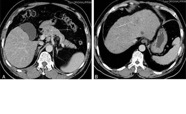
Of special interest is a recent study published by Johnbeck et al. comparing 64Cu-DOTATATE and 68Ga-DOTATOC PET in 59 NET patients on a head-to-head basis []. On a patient level, 64Cu-DOTATATE and 68Ga-DOTATOC performed equally well. However, 64Cu-DOTATATE detected significantly more additional true-positive lesions, confirmed by at least 30 months of follow-up, than 68Ga-DOTATOC (33 versus 7). The authors attributed this difference in lesion detection rate to the shorter positron range of copper-64, with consequent higher spatial resolution and less partial volume effect, rather than to the use of a different peptide. Figure 4 shows an example of a patient where more lesions are seen in the intestinal region with 64Cu-DOTATATE than with 68Ga-DOTATOC. Tumor-to-background ratios as a measure for image contrast were not significantly different between the two tracers. Although the radiation burden of 64Cu-DOTATATE is higher than that of 68Ga-DOTATOC (5.7-8.9 mSv vs. 2.8-4.6 mSv), the use of 64Cu-DOTATATE offers several advantages for use in routine clinical practices associated to the half-life of copper-64, such as supply to peripheral sites from a central production unit and a more flexible scanning window ranging from one hour to at least three hours after injection. A preclinical evaluation of 64Cu-DOTATOC has been published [], but to our knowledge no subsequent clinical studies have been performed.

An external file that holds a picture, illustration, etc.
Object name is ajnmmi0008-0311-f4.jpg

PET/CT (left) and PET (right) scans with 68Ga-DOTATOC and 64Cu-DOTATATE of a patient with intestinal NET and multiple metastases. Additional lesions are seen in the intestinal region with 64Cu-DOTATATE. This research was originally published in JNM. Johnbeck CB, Knigge U, Loft A, Berthelsen AK, Mortensen J, Oturai P, Langer SW, Elema DR and Kjaer A. Head-to-head comparison of 64Cu-DOTATATE and 68Ga-DOTATOC PET/CT: a prospective study of 59 patients with neuroendocrine tumors. J Nucl Med. 2017;58:451-457. © SNMMI.

Copper-64 has also been successfully coupled to TATE by means of a new bifunctional chelator, 5-(8-methyl3,6,10,13,16,19-hexaaza-bicyclo[6.6.6]icosan-1-ylamino)-5-oxopentanoic acid (MeCOSar), forming 64Cu-SARTATE []. Initial preclinical results are promising showing a high uptake in SSTR2-positive tumors []. Further preclinical and clinical studies on this new radiopharmaceutical are ongoing (Australian New Zealand Clinical Trials Registry (ANZCTR) identifier: ACTRN12615000727549) [].

Fluorine-18

Among β+-emitting radioisotopes, fluorine-18 is the most commonly used PET radionuclide in clinical practice and offers several logistic and physical advantages over gallium-68. Large amounts of fluorine-18 activity (> 370 GBq) can be produced with a cyclotron and the half-life (109.8 minutes) is long enough to allow transport to remote hospitals without an on-site cyclotron and it is short enough to avoid extended irradiation of patients. Furthermore, it predominantly decays by positron emission (96.9%) with a low positron energy (Emean = 0.25 MeV) leading to a short positron range (Rmean = 0.6 mm) [].

Meisetschläger et al. evaluated the fluorine-18 labeled SSA, Gluc-Lys-(18F-fluoropropionyl)-Lys-Tyr3-octreotate (Gluc-Lys-[18F]FP-TOCA) (see Figure 1), in 25 patients with SSTR-positive tumors seen on 111In-DTPA-octreotide scan and performed a direct comparison in 16 of these patients []. Gluc-Lys-[18F]FP-TOCA showed a fast and high tumor uptake and was rapidly cleared from the blood, mainly through the kidneys. Tumor uptake reached a plateau at about 40 minutes after injection. In contrast to SSAs labeled with radiometals whose fragments remain trapped after cellular internalization, no such trapping has been observed for SSAs-based radiopharmaceuticals labeled with fluorine-18 such as Gluc-Lys-[18F]FP-TOCA [,]. Nevertheless, more than twice as many lesions were observed with Gluc-Lys-[18F]FP-TOCA than with 111In-DTPA-octreotide. However, a serious impediment to the implementation of Gluc-Lys-[18F]FP-TOCA in routine clinical practice is its time-consuming multistep synthesis with limited radiochemical yield (20%-30%) []. Gluc-Lys-[18F]FP-TOCA has been applied in a few small clinical studies [-], but to our knowledge no further large clinical trials have been performed.

18F-fluoroethyl-triazole-Tyr3-octreotate (18F-FET-βAG-TOCA) (see Figure 1) represents an alternative 18F-octreotate radioligand with a more practical and shorter synthesis route and reasonable radiochemical yield []. Following the promising results in preclinical models [], Dubash et al. carried out a first-in-human study in nine NET patients evaluating the biodistribution and dosimetry of 18F-FET-βAG-TOCA []. The tracer showed a rapid blood clearance with both renal and biliary elimination, whereas 68Ga-DOTA-peptides are mainly eliminated through the kidneys. As such, the highest absorbed dose was received by the gallbladder. Overall, the dosimetry of 18F-FET-βAG-TOCA was similar to other fluorine-18 labeled tracers. Tumor-to-background ratios were high and comparable to values reported for 68Ga-DOTA-peptide PET, resulting in images with excellent contrast. Larger clinical trials for this promising tracer, including a direct comparison with 68Ga-DOTATATE PET/CT are currently ongoing [].

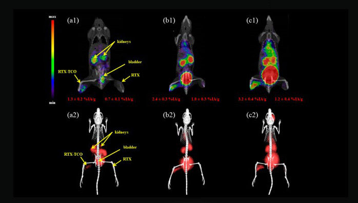
Another fluorine-18 based SSA that is subject of recently initiated clinical trials in NET patients is Al18F-1,4,7-triazacyclononane-1,4,7-triacetate-octreotide (Al18F-NOTA-octreotide) (see Figure 1) (clinicaltrials.gov identifier: NCT03511768). The Al18F-labeling method developed by McBride et al. combines the advantages of a chelator-based radiolabeling method with the unique properties of the radionuclide of choice, fluorine-18 []. In this method, fluorine is firmly bound to Al3+ forming [18F]AlF which is then complexed by a suitable chelator, conjugated to a vector molecule of interest []. Al18F-NOTA-octreotide was developed by Laverman et al. and has been compared to 111In-DTPA-octreotide and 68Ga-NOTA-octreotide in preclinical models [,]. Al18F-NOTA-octreotide proved to have the highest in vitro binding affinity for the SSTR (see Table 2), while a biodistribution in AR42J tumor-bearing mice showed that both tumor uptake and pharmacokinetics were similar with an excellent in vitro and in vivo stability.

Other promising fluorine-18 based tracers for SSTR imaging identified in preclinical studies include 18F-silicon-fluoride-acceptor (SiFA) and 18F-SiFAlin octreotate derivatives [-], 18F-trifluoroborate octreotate (18F-AMBF3-TATE) [] and 18F-fluoroglycosylated octreotate (18F-FGlc-TATE) [].

Scandium-44

Scandium-44 has more recently emerged as a promising radionuclide for PET imaging. There are several methods to produce the radionuclide, for instance by means of a 44Ti/44Sc generator, or using a cyclotron allowing to produce higher quantities [,,]. Scandium-44 mainly decays through positron emission (94.3%) with a somewhat lower positron energy than gallium-68 (Emean = 0.63 MeV) and accordingly lower positron range (Rmean = 2.4 mm). Its half-life of 3.97 hours is convenient for centralized production and distribution []. Even more promising is its use in theranostic applications. Although very similar to gallium(III)-68, the chemical behavior of scandium(III)-44 even more closely resembles that of the therapeutic radiometals, such as lutetium-177 and yttrium-90 []. Therefore, scandium-44 may represent an attractive alternative to gallium-68 for imaging and dosimetry prior to lutetium-177 based therapy []. However, the need for pre-therapy dosimetry has diminished due to the favorable results of the NETTER-1 trial. Moreover, since the half-life of scandium-44 is still limited compared to lutetium-177 (3.97 hours vs. 6.65 days) evaluation at time points later than three days is not possible. With the isotope scandium-47 (100% β- emission), scandium-44 potentially also possesses a true therapeutic match, although research in this area is still in its infancy [,].

Several preclinical studies have been published on various scandium labeled SSAs, such as 44Sc-DOTATOC [,], natSc-DOTATATE [], 44Sc-DOTANOC [,] and 44Sc-1,4,7-triazacyclononane, 1-glutaric acid-4,7-acetic acid(NODAGA)-NOC [].

Rösch et al. performed a clinical proof-of-principle study using generator-produced 44Sc-DOTATOC [,]. High quality PET images of a patient with SSTR-positive liver metastases were acquired at early time points and up to 18 hours after injection. Singh et al. published a proof-of-concept study using cyclotron-produced 44Sc-DOTATOC in two patients with metastatic NET []. Interestingly, scandium-44 was produced at the cyclotron facility at the Paul Sherrer Institut (PSI) in Switzerland and subsequently shipped over 600 km to Zentralklinik Bad Berka (ZBB) in Germany, requiring two and a half half-lives. Eight PET/CT scans were performed at various time points up to 23.5 hours after injection. Excellent tumor uptake was observed with increasing tumor-to-background values over the first four hours after injection. Further clinical studies in larger patient cohorts are scheduled.

Terbium-152

In light of theranostic applications, terbium gained the interest of researchers due to its four medically relevant radioisotopes: terbium-152 and terbium-155 for PET and SPECT imaging, respectively, and terbium-161 and terbium-149 for β- and α-therapy, respectively [,]. The latter also offers the possibility of PET imaging, as demonstrated in a preclinical study evaluating 149Tb-DOTANOC in AR42J tumor-bearing mice []. Terbium-152, suited for diagnostic PET imaging, has a half-life of 17.5 hours and a positron branching ratio of 20.3% with a relatively high positron energy (Emean = 1.14 MeV) and thus higher positron range (Rmean = 5.1) than gallium-68. Terbium-152 - as well as terbium-149 and -155 - can be produced by high-energy proton-induced spallation in tantalum foil targets []. Just like lutetium-177, it belongs to the group of radiolanthanides. Therefore, it can be stably coupled to the chelator DOTA and used for radiolabeling of SSAs []. In a preclinical study in AR42J tumor-bearing mice, the biodistribution of 152Tb-DOTANOC was found to be in good agreement with that of 177Lu-DOTANOC []. These findings suggest that terbium-152 could serve as a theranostic agent for lutetium-177 based therapy. A clinical proof-of-concept was published by Baum et al. in 2017 using 152Tb-DOTATOC in a patient with well-differentiated metastatic NET of the terminal ileum []. Terbium-152 was produced at the Isotope mass Separator On-Line (ISOLDE) facility in CERN in Switzerland, shipped to PSI for separation from the collection matrix and quality control and finally transported to ZBB in Germany for radiolabeling. PET/CT images were acquired at various time points up to 24 hours after injection. All known tumor lesions, visualized on a previous 68Ga-DOTATOC PET scan, were clearly identified. Images were noisier, compared to this previous gallium-68 based PET. This was attributed to the lack of prompt γ-correction by the PET software. Even at 24 hours after injection, increased uptake was observed in several metastases. The authors concluded that the longer half-life of terbium-152, as compared to gallium-68, enabling imaging at later time-points, makes terbium-152 particularly valuable for dosimetry prior to radionuclide therapy []. However, the production of terbium-152 is challenging and currently imposes an important constraint on its implementation in routine clinical practice.

Vector

Developments in SSTR PET radiopharmaceuticals do not only focus on the choice of radionuclide but also on the characteristics of the vector molecule. The radiopharmaceuticals described above contain the somatostatin receptor agonist octreotide, or an analog of octreotide, as vector molecule, with a predominance of TOC and TATE. Improved tumor targeting may be achieved for instance by using vector molecules having a higher binding affinity for the SSTR or a broader affinity profile for the different receptor subtypes or by using compounds recognizing a higher number of binding sites, such as the SSTR antagonists.

Somatostatin receptor agonists

Many tumor types that show SSTR expression, predominantly express SSTR2 [], which makes it the main target for the development of SSTR ligands for imaging and therapy. However, as mentioned above, a wide variability in subtype expression has been observed across and within different tumor types []. Therefore, there is great interest in SSTR ligands with a broader affinity profile to increase tumor uptake and to expand the number of tumors eligible for SSTR imaging []. Of the 68Ga-DOTA-peptides currently used in clinical practice, 68Ga-DOTANOC has the widest affinity profile with high affinities for SSTR2, SSTR5 and to a lesser extent SSTR3 []. However, as discussed above, there is no conclusive evidence to prefer this ligand in clinical practice and large trials are warranted [], but properly powered prospective trials testing this hypothesis will probably not be available for a long time. Another SSA that has been evaluated early on in clinical trials is the long-acting SSA lanreotide (see Figure 1). Coupled to the chelator DOTA, DOTA-lanreotide (DOTALAN) and the radiolabeled compounds 111In/90Y-DOTALAN showed a high affinity for SSTR subtype 2-5 []. However, this was only confirmed for SSTR2 and 5 by Reubi et al. []. Following promising clinical results with 111In-DOTALAN [,], DOTALAN was labeled with gallium-68 to allow PET imaging. After an initial positive evaluation of 68Ga-DOTALAN in 11 patients with lung cancer (three small cell and three non-small cell lung cancer) or thyroid cancer (two medullary and three radioiodine negative thyroid cancer) [], the tracer was used to identify patients who might benefit from PRRT with 90Y-DOTALAN in a group of NET patients not qualified for PRRT with 68Ga-DOTATOC despite progressive disease []. Tumor-to-background ratios were significantly higher for 68Ga-DOTATOC and more tumor sites (106 vs. 53) were detected with 68Ga-DOTATOC than with 68Ga-DOTALAN. Demirci et al. compared 68Ga-DOTALAN with 68Ga-DOTATATE in a group of 11 NET patients and one patient with meningioma. 68Ga-DOTATATE performed better than 68Ga-DOTALAN with a significantly higher lesion uptake and higher lesion detection rate (63 vs. 23 out of a total of 67 tumor lesions detected with both tracers) []. Traub-Weidinger et al. evaluated the SSTR status in a heterogeneous group of thyroid cancer patients with progressive disease using two tracers with a distinct SSTR subtype affinity profile, 68Ga-DOTALAN and 68Ga-DOTATOC, in 28 patients []. On a patient basis, 12 patients were negative with both SSTR tracers, while mixed results were observed in three patients (two patients negative with 68Ga-DOTALAN, but positive with 68Ga-DOTATOC and one patient vice versa). On a region-based analysis half of the 38 regions positive on SSTR imaging (out of a total of 196 regions) showed mixed results. The authors concluded that, due to the heterogeneous SSTR profile of thyroid cancer lesions, patients with progressive disease may benefit from imaging with different SSTR PET tracers for individualized targeted therapy stratification.

In a first attempt to develop an actual pansomatostatin radiopharmaceutical with high affinity for all SSTR subtypes, the cyclooctapeptide KE108 with pansomatostatin characteristics was modified to couple the chelator DOTA (DOTA-D-Dab-Arg-Phe-Phe-D-Trp-Lys-Thr-Phe or KE88) and allow subsequent radiolabeling with indium-111 and gallium-68 []. Although the resulting tracers were able to bind all SSTR subtypes with high affinity, in vitro internalization of the ligand-receptor complex for the SSTR2 was low compared to the SSTR3 and this was reflected by a low in vivo uptake observed in SSTR2-expressing tumors and fast wash out []. In another preclinical study, Fani et al. synthesized and evaluated several bicyclic somatostatin-based analogs of which DOTA-Tyr-cyclo(DAB-Arg-cyclo(Cys-Phe-D-Trp-Lys-Thr-Cys)) or AM3, showing a high affinity for SSTR2, SSTR3 and SSTR5, was the most promising []. Efficient background clearance and high tumor uptake with 68Ga-AM3 were observed in SSTR2 tumor-bearing mice. Tatsi et al. used the native peptide hormone somatostatin-14 (SS14) as a basis to develop two SS14-derived analogs with a high affinity for all SSTR subtypes and label them with indium-111 []. However, both tracers showed poor in vivo stability. Similarly, Maina et al. used the more stable native peptide somatostatin-28 (SS28) as a basis for the development of DOTA-Ser,Leu,D-Trp,Tyr-SS28 (DOTA-LTT-SS28) and the radioligand 111In-DOTA-LTT-SS28 []. The compounds displayed a high affinity for all SSTR subtypes and triggered internalization of SSTR2, SSTR3 and SSTR5. 111In-DOTA-LTT-SS28 showed a high and specific uptake in SSTR2-, SSTR3- and SSTR5-expressing xenografts in mice and a much higher in vivo stability than the SS14-derived tracers developed by Tatsi et al. The authors concluded that 111In-DOTA-LTT-SS28 is the first true (pan)somatostatin radioligand and may serve as a model for the further development of pansomatostatin radioligands []. Very recently, Liu et al. reported the development of gallium-68 labeled pasireotide (SOM230 or PA1), a longacting synthetic SSA with a high affinity for SSTR1, SSTR2, SSTR3 and SSTR5 [,]. The resulting radiotracer 68Ga-DOTA-PA1 displayed a significantly higher in vitro uptake than 68Ga-DOTATATE in three human lung cancer cell lines: lung adenocarcinoma (A549), lung squamous carcinoma (H520) and pulmonary giant cell carcinoma (PG). PET images of A549 tumor-bearing mice showed a high tumor uptake and better signal-to-noise ratio with 68Ga-DOTA-PA1 than with 68Ga-DOTATATE and the PET signal correlated with the total expression of SSTRs and not only SSTR2, as determined by Western blotting. The authors concluded that 68Ga-DOTA-PA1 and its analogs may hold potential for SSTR imaging in clinical practice, especially lung tumors [].

Therapeutic purposes have also driven some new developments in the vector part of SSTR ligands aiming for higher tumor retention to improve therapeutic efficiency and preferably lower kidney dose to reduce PRRT toxicity. A recent example is given by Tian et al. who conjugated an Evans blue (EB) analog onto octreotate which allows reversible binding of EB-TATE to albumin to prolong half-life in blood, resulting in a SSA with long circulation time []. EB-TATE was then labeled with yttrium-90 through the chelator DOTA and injected in AR42J tumor bearing mice. 90Y-DOTA-EB-TATE showed high tumor uptake resulting in a complete regression of the tumors []. These promising results were quickly translated into a first-in-human clinical trial by Zhang et al. evaluating the safety, pharmacokinetics and dosimetry of 177Lu-DOTA-EB-TATE in five NET patients as compared to 177Lu-DOTATATE in three other NET patients []. The new compound was well tolerated without adverse symptoms. Tumor dose was 7.9-fold higher with 177Lu-DOTA-EB-TATE, while the effective dose was not significantly different between 177Lu-DOTA-EB-TATE and 177Lu-DOTATATE. However, kidney and bone marrow dose increased 3.2- and 18.2-fold, respectively [], so this radiopharmaceutical does not offer an improved therapeutic ratio compared to the current radiopharmaceutical of choice, 177Lu-DOTATATE.

Somatostatin receptor antagonists

All SSTR-targeting radiopharmaceuticals described above are somatostatin agonists. After binding to the SSTR, the ligand-receptor complex is usually internalized, allowing tracer metabolites to accumulate in the target cells []. For a long time, it was believed that this process of internalization and subsequent accumulation of the radioligands was essential for high-contrast imaging of SSTR-positive lesions []. However, in 2006, Ginj et al. observed in cell cultures expressing human SSTR2 and SSTR3, that antagonists labeled considerably more receptor sites than agonists []. This was reflected by the significantly higher tumor uptake seen in mice bearing SSTR2 and SSTR3-expressing tumors after injection with the corresponding antagonist as compared to those injected with the agonist []. Especially striking was the fact that counterintuitive to this observation the SSTR2 antagonist, 111In-DOTA-pNO2-Phe-c(D-Cys-Tyr-D-Trp-Lys-Thr-Cys)D-TyrNH2 (111In-DOTA-BASS) (see Figure 1), showed a more than sevenfold lower affinity for the SSTR2 than the SSTR2 agonist 111In-DTPA-TATE in this study (50% inhibitory concentration (IC50) of 9.4 ± 0.4 nM vs. 1.3 ± 0.2 nM) []. In 2011, Wild et al. published a pilot study in five patients, one metastatic follicular thyroid carcinoma and four NETs, evaluating the biodistribution, tumor uptake and detection of tumor lesions with the SSTR antagonist 111In-DOTA-BASS in a head-to-head comparison with 111In-DTPA-octreotide []. Tumor uptake was up to four times higher with the antagonist, while renal, liver and spleen uptake were lower. 111In-DOTA-BASS detected more lesions than 111In-DTPA-octreotide (25 vs. 17 out of 28). The three missed bone lesions were negative on the 111In-DTPA-octreotide scan as well.

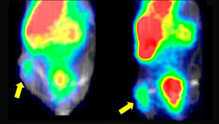
Further advances in the preclinical setting in search of more potent SSTR2 antagonists labeled with PET radioisotopes led to the identification of Cpa-c[D-Cys-Aph(Hor)D-Aph(Cbm)-Lys-Thr-Cys]-D-Tyr-NH2 (JR11) (see Figure 1) as a promising compound for implementation in the clinical field []. Based on the results of previous affinity studies and an in vivo biodistribution study [], 68Ga-NODAGA-JR11 (68Ga-OPS202) was selected for a first clinical evaluation by Nicolas et al. [,]. 12 patients with GEP NET and a positive 68Ga-DOTATOC PET/CT scan in the previous six months were administered two microdoses of 68Ga-NODAGA-JR11 with ascending peptide masses at different study visits and underwent subsequent PET/CT scans. 68Ga-NODAGA-JR11 showed a fast blood clearance and favorable biodistribution with both peptide doses as compared to 68Ga-DOTATOC (lower hepatic, pancreatic, gastro-intestinal and splenic uptake with 68Ga-NODAGA-JR11) [,]. This was reflected by higher tumor-to-background ratios observed with 68Ga-NODAGA-JR11 and further translates to a significantly higher number of detected tumor lesions and higher lesion-based overall sensitivity (94% and 88% for 50 µg and 15 µg 68Ga-NODAGA-JR11, respectively, vs. 59% for 68Ga-DOTATOC) []. An example is shown in Figure 5. The effective dose is in line with 68Ga-DOTA-peptides (24 ± 2 µSv/MBq for 68Ga-NODAGA-JR11 vs. 21 ± 3 µSv/MBq for 68Ga-DOTATATE and 68Ga-DOTATOC []), although some differences in organ doses are observed due to a slightly different biodistribution []. Overall, 68Ga-NODAGA-JR11 was well tolerated []. However, SSTR antagonists could possibly counteract the effects of SSAs, which may be important in patients with functioning NETs []. Therefore, caution is required in anticipation of more safety data []. Currently a multicenter clinical trial evaluating the optimal dose and safety of 68Ga-NODAGA-JR11 for PET imaging is ongoing (clinicaltrials.gov identifier: NCT03220217).

An external file that holds a picture, illustration, etc.
Object name is ajnmmi0008-0311-f5.jpg

Maximal-intensity projections (A and C) and PET/CT (B and D) scans with 68Ga-NODAGA-JR11 (A and B) and 68Ga-DOTATOC of a patient with ileal NET and bilobar liver metastases. Liver magnetic resonance imaging (MRI) was performed four months later, with delayed post-contrast acquisitions (E) and diffusion-weighted images (F), confirming the additional metastases missed or questionable (arrow with question mark) with 68Ga-DOTATOC. Note the lower background activity in the liver, intestine and thyroid with 68Ga-NODAGA-JR11. Dashed lines in (A and C) denote the level of transversal slices in (B, D, E and F). This research was originally published in JNM. Nicolas GP, Schreiter N, Kaul F, Uiters J, Bouterfa H, Kaufmann J, Erlanger TE, Cathomas R, Christ E, Fani M and Wild D. Sensitivity comparison of 68Ga-OPS202 and 68Ga-DOTATOC PET/CT in patients with gastroenteropancreatic neuroendocrine tumors: a prospective phase II imaging study. J Nucl Med. 2018;59:915-921. © SNMMI.

The higher tumor uptake achieved with SSTR antagonists may also prove useful for therapeutic purposes. Preclinical studies comparing 177Lu-DOTA-JR11 (177Lu-OPS201) with 177Lu-DOTATATE observed higher tumor uptake and longer residence times, resulting in higher tumor doses delivered by the antagonist as compared to the agonist [,]. In a clinical pilot study by Wild et al. in four patients with progressive NETs, more than threefold higher tumor doses and twofold higher tumor-to-kidney and tumor-to-bone marrow dose ratios were observed using a test dose of 177Lu-DOTA-JR11 as compared to 177Lu-DOTATATE []. All patients were subsequently treated with 177Lu-DOTA-JR11, resulting in partial remission in two patients, mixed response in one patient and stable disease in the last patient, and as such proving the clinical feasibility of PRRT using radiolabeled SSTR antagonists. Currently, a multicenter clinical trial evaluating the safety and efficacy of 177Lu-DOTA-JR11 (clinicaltrials.gov identifier: NCT02592707) as well as a study evaluating the theranostic couple 68Ga-DOTA-JR11 and 177Lu-DOTA-JR11 (clinicaltrials.gov identifier: NCT02609737) are ongoing.

Radiolabeled SSTR antagonists might also prove to be especially useful for imaging and therapy of cancer types with a typically lower SSTR expression such as breast cancer []. Several preclinical studies observed enhanced tumor targeting in various human SSTR2-expressing tumor samples, including breast carcinoma, by means of in vitro autoradiography using an SSTR2 antagonist in comparison to the SSTR2 agonist [-]. This finding was not confirmed in a recent preclinical study by Dude et al. using the human luminal breast cancer model, ZR-75-1, with endogenous SSTR2 expression and negligible expression of other SSTR subtypes, where 68Ga-NODAGA-JR11 had a lower tumor uptake than 68Ga-DOTATOC and 68Ga-DOTATATE []. This was tentatively explained by the authors by the fact that they used an endogenously expressing cell line, which may have a lower amount of low-affinity, antagonist-specific binding sites. Interestingly, although 68Ga-DOTATATE has a higher affinity for the SSTR2 than 68Ga-DOTATOC, the latter was found to have the highest tumor uptake. Additional studies are warranted to further investigate the role of SSTR2 antagonists in breast cancer imaging [].

Conclusion

Advances in SSTR PET ligands occur on two major fronts: the radionuclide and the peptide vector. Other radionuclides could offer a solution to practical, economical and regulatory barriers to the adoption of 68Ga-DOTA-peptide PET, with additional physical advantages such as lower positron range and longer half-life. Developments concerning peptide vectors are mainly driven by the need for improved lesion targeting, especially for tumors with low SSTR expression. Therefore, advances on both fronts are largely complementary. Several promising new PET ligands for clinical SSTR imaging are currently in the pipeline and good results have been demonstrated in phase II trials. Clinical adoption in the near future is a realistic scenario.

Acknowledgements

This research was funded by the project from “Kom op tegen Kanker”: “PET/MR imaging of the norepinephrine transporter and somatostatin receptor in neural crest and neuroendocrine tumors for better radionuclide therapy selection” and received support from Research Foundation-Flanders (FWO) (G0D8817N). Frederik Cleeren is a Postdoctoral Fellow of FWO (12R3119N). Christophe M. Deroose is a Senior Clinical Investigator at the FWO.

Disclosure of conflict of interest

Christophe M. Deroose received grants and personal fees from Novartis, Terumo, AAA, Ipsen, Sirtex, Bayer outside the submitted work.

The information comes from:
https://www.ncbi.nlm.nih.gov/pmc/articles/PMC6261874/


Development of a Potential Gallium-68-Labelled Radiotracer Based on DOTA-Curcumin for Colon-Rectal Carcinoma: From Synthesis to In Vivo Studies

Abstract

Colorectal cancer is the third most commonly occurring cancer in men and the second most commonly occurring cancer in women worldwide. We have recently reported that curcuminoid complexes labelled with gallium-68 have demonstrated preferential uptake in HT29 colorectal cancer and K562 lymphoma cell lines compared to normal human lymphocytes. In the present study, we report a new gallium-68-labelled curcumin derivative (68Ga-DOTA-C21) and its initial validation as marker for early detection of colorectal cancer. The precursor and non-radioactive complexes were synthesized and deeply characterized by analytical methods then the curcuminoid was radiolabelled with gallium-68. The in vitro stability, cell uptake, internalization and efflux properties of the probe were studied in HT29 cells, and the in vivo targeting ability and biodistribution were investigated in mice bearing HT29 subcutaneous tumour model. 68Ga-DOTA-C21 exhibits decent stability (57 ± 3% after 120 min of incubation) in physiological media and a curcumin-mediated cellular accumulation in colorectal cancer cell line (121 ± 4 KBq of radiotracer per mg of protein within 60 min of incubation). In HT29 tumour-bearing mice, the tumour uptake of 68Ga-DOTA-C21 is 3.57 ± 0.3% of the injected dose per gram of tissue after 90 min post injection with a tumour to muscle ratio of 2.2 ± 0.2. High amount of activity (12.73 ± 1.9% ID/g) is recorded in blood and significant uptake of the radiotracer occurs in the intestine (13.56 ± 3.3% ID/g), lungs (8.42 ± 0.8% ID/g), liver (5.81 ± 0.5% ID/g) and heart (4.70 ± 0.4% ID/g). Further studies are needed to understand the mechanism of accumulation and clearance; however, 68Ga-DOTA-C21 provides a productive base-structure to develop further radiotracers for imaging of colorectal cancer.

1. Introduction

Colorectal cancer (CRC) is the third most commonly occurring cancer in men and the second in women worldwide, with more than 1.4 million new cancer cases every year []. India has a relatively low prevalence of CRC in comparison to other countries, mainly attributed to differences in dietary patterns and lifestyles []. It is widely known that curcumin (1,7-bis(4-hydroxy-3-methoxyphenyl)-1,6-heptadiene-3,5-dione), a phyto-polyphenol pigment isolated from the dried rhizomes of Curcuma longa L., has been used for centuries in Asian countries as a dietary spice and traditional drug to treat many different diseases []. Besides having antioxidant, anti-inflammatory, anti-bacterial and anti-amyloid properties, curcumin has been shown also to possess anti-cancer activities [,]. In particular, the ability to inhibit proliferation of a wide variety of tumour types, including colorectal cancer, has been highlighted by recent studies in vitro and in vivo [,,]. Several studies suggest that curcumin changes the gene expression profiles and signalling pathways such as those involving COX-2 enzymes as well as NF-κB, cyclin D1 and p53 proteins [,,]. Although the mechanism of curcumin’s high accumulation and cytotoxic activity against HeLa, MCF-7, HT29, and HCT-116 cancer cell lines are still unclear [,], curcumin itself is a promising tumour-targeting moiety for the development of early diagnosis and therapeutic agents. Nuclear medicine imaging provides information about biological processes occurring at a molecular level in vivo; this occurs by following the fate of radiolabelled compounds. Such radiotracers can be substrates for metabolic pathways overexploited in tumour cells or have particular affinity for receptors overexpressed in tumours. Hence, curcumin derivatives labelled with proper β+ emitter radionuclides or curcumin-based radio-metal complexes have the potential to be diagnostic tools for positron emission tomography (PET). Currently, radiolabelled curcuminoids have been mostly investigated for imaging of the central nervous system (CNS) where several curcumin-based probes labelled with fluorine-18 have demonstrated promise in the early detection of Alzheimer’s disease [,]. In oncology, curcuminoids as imaging agents are underinvestigated. We have recently reported that curcuminoid complexes labelled with gallium-68 demonstrated preferential uptake in HT29 colorectal cancer and K562 lymphoma cell lines compare to normal human lymphocytes. In that study, the core radio-metal was directly linked to two curcuminoid molecules through the keto-enol moiety [,]. The curcuminoid complexes showed good stability in PBS and human serum but were rapidly degraded in whole human blood. These findings, in addition to the low stability and solubility of curcumin structures in physiological media [], limited the possibility to further study these kind of compounds in vivo. In the present study, we report a new gallium-68-labelled curcumin derivative with curcumin linked to an efficient gallium-68 chelator, 1,4,7,10-tetraazacyclododecane-1,4,7,10-tetraacetic acid (DOTA), and its initial validation as a marker for early detection of CRC. The synthetic pathway, chemical characterization, labelling methods, stability, cell uptake, internalization and efflux studies on HT29 cells and in vivo micro-PET imaging and biodistribution in colorectal tumour bearing mice of this radiotracer are herein reported.

2. Results

2.1. Synthesis and Chemical Characterization

Synthesis of 1,4,7,10-tetraazacyclododecane-1,4,7,10-tetraacetic(1,7-bis(4-hydroxy-3-methoxyphenyl)-1,6-heptadiene-3,5-dione (DOTA-C21) was carried out in two steps as shown in Figure 1. The curcumin-based conjugate was identified and completely characterized by ESI-LC/MS (Figure S1) and 1H-,13C-NMR spectroscopy (Figures S2, S3). ESI-LC/MS spectrum exhibits two isomeric peaks (m/z = 798.3; [M + H]+) that were attributed to contemporary presence of two main isomeric forms (diketo and keto-enol) as previously reported for curcumin and its derivatives [,]. Gallium complexes were obtained by reacting a DOTA-C21 solution with a Ga(NO3)3 solution at 95 °C for 30 min in a 1.5:1 metal to ligand molar ratio using a 0.4 M ammonium acetate solution as buffer. Complexes formation was identified by ESI-LC/MS analysis thanks to the typical isotopic pattern of gallium ion (Figures S4, S5). Similar to the free ligand analysis, the spectrum exhibits two compounds with the same m/z (retention times: 2.8 and 3.3 min, respectively) both corresponding to Ga-DOTA-C21 complexes (m/z = 864.2 [M69Ga + H]+ – 866.2 [M71Ga + H]+). To elucidate chelation mode and binding sites, 1H-NMR titration of DOTA-C21 with Ga3 + was also performed (Figure S6) and the gallium(III)-complex was characterized by NMR (Figure 2, Figure S7) as well as LC-MS/MS fragmentation experiments (Figure 3, Figure S8).

An external file that holds a picture, illustration, etc.
Object name is molecules-24-00644-g001.jpg

Synthetic pathway for the preparation of DOTA-C21 and its complexation with Ga(III) by reacting DOTA-C21 with Ga(NO3)3 at 1.5:1 metal-to-ligand molar ratio in 0.4 M ammonium acetate solution for 30 min at 95 °C.

An external file that holds a picture, illustration, etc.
Object name is molecules-24-00644-g002.jpg

Aliphatic region overlap of 1H-, 13C-HSQC-NMR spectra of DOTA-C21 (red (CH2) and purple (CH/CH3)) and Ga-DOTA-C21 (green (CH2) and blue (CH/CH3)). All the spectra were acquired in MeOD-d4 at 25 °C ([DOTA-C21] = 0.63 mM). Atom numbering refers to Figures 11 and 12.

An external file that holds a picture, illustration, etc.
Object name is molecules-24-00644-g003.jpg

LC-MS/MS fragmentation experiments on Ga-DOTA-C21 complexes with m/z 864.5 [M + H]+. HPLC-MS chromatogram (A). Fragmentation pathway of the m/z 864.5 [M + H]+ ion corresponding to the peak with retention time 5.9 min (B). Fragmentation pathway of the m/z 864.5 [M + H]+ ion corresponding to the peak with retention time 6.5 min (C).

1H- and 13C-NMR spectra displayed a strong shift of both protons and carbon with respect to the free ligand when gallium(III) is added. In particular, methylene protons of the chelator ring, briefly r1 and r2, are no longer chemical shift equivalent since the interconversion between pseudo-axial (Ha) and pseudo-equatorial (He) conformation is prevented by metal complexation. Geminal protons Ha(r1) and He(r1) appear as broad triplets at 4.00 ppm and 3.38, respectively, due to similar values of geminal (2JHH) and vicinal (3JHH) coupling constants (~14Hz). The same situation occurs for Ha(r2) and He(r2) that fall at 3.47 ppm and 3.41 ppm, respectively. Assignments of ring protons was confirmed by 1H,13C-HSQC spectrum, Ha/He(r1) show cross-peaks with carbon at 54.65 ppm (C(r1)) and Ha/He(r2) with a carbon at 57.11 ppm (C(r2)). The methylene groups of acetate arms are not chemical shift equivalent in gallium complex and appear as broad singlets in proton spectrum. The following reasonable assignments can be proposed (for atom numbering refers to Section 4.1.1 and Section 4.1.2): CH2 in α to amide and nitrogen (atom numbering 15) 1H- 3.79 ppm and 13C- 60.64 ppm; CH2 in α to carboxylic group involved in gallium coordination (atom numbering 24 and 26) 1H- 3.83 ppm and 13C- 59.41 ppm; CH2 in α to free carboxylic group (atom numbering 25) 1H- 3.74 ppm, 13C- 61.68 ppm.

Moreover, UV absorption and fluorescent emission spectra upon increasing addition of Ga3+ from free ligand were acquired (Figures S9, S10). These analyses showed that DOTA-C21 displays an absorption maximum at 410 nm, typical of curcumin structure in the keto-enol form. Upon addition of gallium (III) solution, an increase in absorbance is observed at 290 nm, while no changes are observed for the absorption maximum. Fluorescence emission spectra of DOTA-C21 (λex 410 nm) do not show any significant change after gallium(III) addition.

2.2. Radiolabelling of the Chelator-Curcuminoid Derivatives with Gallium-68

Radiolabelling was performed using two methods. In the first, postprocessing of the generator elution was performed by cation exchange purification [] and kinetics of the labelling were studied between 1 and 20 min by TLC. Kinetics were fast and the incorporation yield was 97 ± 2% after 5 min at 95 °C (n = 3). Postprocessing of the generator elution minimizes the metal cation impurities allowing a complete radiolabelling of DOTA-C21 by using only 10 nmol of the precursor. Representative TLC analyses are shown in Figure S11. In the second method, labelling without processing of the elution was attempted with increasing amount of precursor (20, 40, 60, 80 nmol). In this case, a reproducible radiolabelling could only be achieved with 80 nmol of DOTA-C21 after 10 min at 95 °C, with an incorporation yield ranging from 60 to 80%. The crude mixture was further purified by SPE to eliminate unreacted gallium-68 and hydrolysed products. Radiochemical purity was assessed by UHPLC (RCP > 95%, RCY = 55 ± 5%, n = 10). Representative UHPLC chromatograms (radiochemical detector) of a 68Ga-DOTA-C21 preparation before and after SPE purification are shown in Figure 4. Similarly, to the complexation reaction with natural gallium, two main radioactive species were obtained with a retention time of 4.7 and 5.1 min, respectively. Based on the data gathered for the non-radioactive preparation, the peaks were assigned to two 68Ga-DOTA-C21 isomeric complexes. A further peak at 5.3 min was recorded (ca. 4% of total radioactivity) and was likely due to a gallium-68 complexes with a degraded form of the precursor at labelling conditions. In both methods, the pH was kept constant around 4.

An external file that holds a picture, illustration, etc.
Object name is molecules-24-00644-g004.jpg

Representative UHPLC chromatograms (radiochemical detector) of a 68Ga-DOTA-C21 preparation before (A) and after (B) SPE purification. (1) 68Ga-free, (2) 68Ga-hydrolyzed products, (3) 68Ga-labelled byproducts, (4) and (5) 68Ga-DOTA-C21 isomers, (6) 68Ga-labelled degradation product.

2.3. In Vitro Stability Studies

In vitro stability of 68Ga-DOTA-C21 complexes in PBS, human serum (HS) and whole human blood (HB) were determined according to peak integration of analytical UHPLC (Figure 5). Stability of 68Ga-DOTA-C21 in PBS was more than 88 ± 3% after 120 min at 37 °C. In HS, the radiotracer was also stable with 85 ± 3% of intact compound after 120 min of incubation. On the contrary, in whole blood, 68Ga-DOTA-C21 was partially degraded to more polar metabolites already after 10 min of incubation. The amount of intact compound ranged from 74 ± 2% after 10 min to 57 ± 3% after 120 min of incubation.

An external file that holds a picture, illustration, etc.
Object name is molecules-24-00644-g005.jpg

Stability of 68Ga-DOTA-C21 complex incubated over time (10, 40, 70, 120 min.) in different media (n = 3, mean ± SD).

2.4. Uptake, Internalization and Efflux in Colorectal Cancer Cell Line

In vitro 68Ga-DOTA-C21 uptake studies demonstrated time-dependent cellular accumulation in HT29 colorectal cancer cell line. Total uptake increased continuously in the first 60 min of incubation up to 121 ± 4 KBq of radiotracer per mg of protein (n = 3). At 60 min post incubation, 83% of total radioactivity was internalized in the cells. Conversely, 68GaCl3, used as a negative control, demonstrated significantly lower in vitro cell uptake (20 ± 4 KBq/mg of protein after 60 min of incubation, n = 3) confirming the curcuminoid-mediated uptake by HT29 cells (Figure 6A,B). The cellular uptake of 68Ga-DOTA-C21 is comparable to that of the compound obtained by direct labelling of curcumin with gallium-68 (i.e., 68Ga(CUR)2 complexes []), however the internalized fraction was more than doubled. To test for receptor-mediated uptake, HT29 cells were preincubated with a 200-fold excess of calcitriol, the natural ligand for vitamin D receptor (VDR), and then 68Ga-DOTA-C21 was added. The experiments were performed to assess whether 68Ga-DOTA-C21 uptake was mediated by VDR since it has been reported that curcumin has a structure suitable to bind nuclear vitamin D receptor. The difference between uptake with or without calcitriol was not statistically significant (n = 3, Figure 6C) suggesting that the VDR receptor was not involved in the uptake or that the receptor cannot be saturated in these conditions.

An external file that holds a picture, illustration, etc.
Object name is molecules-24-00644-g006.jpg

Uptake and internalization at different time points of 68Ga-DOTA-C21 in HT-29 colorectal cancer cell line (n = 3, mean ± SD) (A). Comparison between the uptake of 68Ga-DOTA-C21 and 68GaCl3 in HT-29 cells indicating curcumin structure dependent accumulation (n = 3, mean ± SD) (B). Comparison between the uptake of 68Ga-DOTA-C21 and 68Ga-DOTA-C21 + calcitriol in HT-29 cells (n = 3, mean ± SD) (C).

For efflux studies, cells were incubated with 68Ga-DOTA-C21 and the uptake was stopped after 60 min by removing the supernatant. Then, cells were washed, fresh medium was added and they were incubated again for different periods of time. Media were collected and radioactivity released from the cells was measured.68Ga-DOTA-C21 showed a slow externalization pattern with around 75% (n = 3) of the radiotracer remaining within the cell after 60 min of incubation (Figure 7).

An external file that holds a picture, illustration, etc.
Object name is molecules-24-00644-g007.jpg

Externalization of 68Ga-DOTA-C21 in HT-29 colorectal cancer cell line at different time points (n = 3, mean ± SD).

2.5. PET Imaging and Biodistribution

HT29 tumour-bearing mice were injected i.p. with 68Ga-DOTA-C21 and PET/CT imaging was taken 1 h and 2 h post injection (Figure 8). The following day, the same cohort of mice was injected i.v. with 68Ga-DOTA-C21 and dynamic PET/CT imaging was registered for the first 10 min to assess initial distribution of the tracer. The 1 h post injection time point was retaken to evaluate potential differences in the biodistribution pathway related to the type of injection. Mice were sacrificed at 1.5 h post injection, and ex-vivo biodistribution performed. As per PET/CT imaging analysis, upon i.v. injection, 68Ga-DOTA-C21 was taken up rapidly in the heart and liver, and began accumulating into the tumour within the first minute of circulation. The tumour was immediately visible over background (muscle). At 1 h post injection, tumour accumulation of the tracer was 2.27 ± 0.85% ID/cc (n = 3) with a tumour to muscle ratio (T/M) of 1.91 ± 0.47 (n = 3). 68Ga-DOTA-C21 appeared to have both renal and hepatic clearance. Radioactivity in the heart, which reflected the blood pool, was surprisingly high at 1 h post injection (5.8 ± 0.7% ID/cc, n = 3). Similar biodistribution was observed when the tracer was delivered i.p. At 1 h post injection, the tumour had an uptake of 1.89 ± 0.45% ID/cc (n = 5) and a T/M of 3.59 ± 2.76 (n = 5). At 2 h post injection, tumour uptake increased to 2.60 ± 0.27% ID/cc (n = 5) which was reflected by an increase in the T/M to 5.41 ± 3.62 (n = 5) (Figure 9 and Figure S12A–C). It is worth noting that, when analysing tumour uptake in each single animal, there was no appreciable variation between 1 h and 2 h post injection in animals with an initial low tumour uptake. On the contrary, animals with a tumour uptake greater than 4% ID/cc after one hour, tended to accumulate more radiotracer (Figure S12D). No correlation was found between T/M ratio and tumour size (Figure S12E). Ex-vivo biodistribution confirmed the described accumulation (Figure 9). Tumour uptake was 3.08 ± 0.53% ID/cc (n = 5) with a T/M of 2.29 ± 0.62 (n = 5). As already observed by PET, clearance occurred via both renal and hepatobiliary excretion, bone uptake was barely detectable over background and no significant radioactivity was seen in the intracranial region, suggesting that 68Ga-DOTA-C21 is unlikely to cross an intact blood–brain barrier. Blood radioactivity was high, suggesting that a high amount of the radiotracer was in circulation or bound to blood constituents such as serum albumin. Further studies evaluating the clearance mechanism of 68Ga-DOTA-C21 are underway to understand the high blood circulation, however one explanation may be that 68Ga-DOTA-C21 pharmacokinetics follow a two-compartment model [,].

An external file that holds a picture, illustration, etc.
Object name is molecules-24-00644-g008.jpg

Representative microcoronal-PET/CT scans of HT29 colorectal tumour bearing mouse at 60 min (A) and 120 min (B) post i.p. injection of ca. 37 MBq of 68Ga-DOTA-C21.

An external file that holds a picture, illustration, etc.
Object name is molecules-24-00644-g009.jpg

Biodistribution in multiple organs of 68Ga-DOTA-C21 in HT-29 colorectal cancer bearing nude mice (n = 5, mean ± SD) at 90 min post i.v. injection. (A). Ratios of the tumour uptake to major organs (n = 5, mean ± SD) for 68Ga-DOTA-C21 at 90 min post i.v. injection (B).

2.6. Metabolism

Mouse blood was drawn 1.25–1.5 h post injection, processed and analysed by radio-HPLC. Out of the two isomers present in the parent compound, one disappears in the blood and only the more polar one (Rt = 7.7 min) persists in all the analysed mice (n = 5) (Figure S13). It is likely that physiological media influence the keto-enol tautomerism of 68Ga-DOTA-C21, stabilizing the diketo form [].

3. Discussion

The use of curcumin and its derivatives as potential therapeutics in many medical applications has been largely attested by a long list of scientific literature [,,]. We previously demonstrated that 68Ga-labelled curcumin complexes had preferential accumulation in HT29 colorectal cancer cells compared to other cancer cell lines and lymphocyte cultures []. However, the stability of these complexes in blood was low as the curcuminoid backbone was directly linked to the radio-metal through the keto-enol moiety, resulting in positively charged 2:1 ligand to metal ratio complexes. The present study is focused on evaluating whether a suitable curcumin derivative that is more stably labeled with gallium-68 provides a radiotracer that preferentially accumulates in CRC lesions, and could be a useful early detection agent which enables nuclear medicine imaging techniques. CRC is globally the third most common type of cancer and is usually diagnosed by invasive techniques like sigmoidoscopy or colonoscopy, during which a colon specimen is collected. These procedures are then followed by medical imaging (usually CT scan) to determine if the disease has spread. While small polyps may be removed during colonoscopy, the cancerous state of large polyps may be assessed by a biopsy []. The introduction of a PET radiotracer that specifically accumulates in CRC might be useful for verifying the nature of these lesions without resort to invasive methods. Moreover, treatments used for colorectal cancer may include some combination of surgery, radiation therapy, chemotherapy and targeted therapy and nuclear medicine imaging may be a suitable technique to stage and assess follow-up of the patients.

In the present study, the structure of the first generation of gallium-68 curcumin complexes has been improved by adding DOTA, a strong gallium-68 chelator. DOTA was linked through an (amino)ethyl spacer to one of the phenol groups of the curcumin backbone. With the addition of DOTA, the new bio-conjugate (namely DOTA-C21) has an enhanced water solubility and a higher stability of the complex with gallium-68 is achieved in physiological conditions. Despite its solubility in water, 1H-NMR spectrum of DOTA-C21 in D2O (Figure S2A) exhibited extremely broad signals in the aromatic region, suggesting that the hydrophobic backbone of curcumin interacted strongly by intramolecular π−π stacking. The signals could be sharpened by reducing the solvent polarity, for example, switching to methanol-d4, which reduced aggregation. The 1H-NMR spectrum in methanol-d4 (Figure S2B) provided signals in the aromatic region attributable to the asymmetric curcumin moiety of DOTA-C21, while methylene signals of the ethyl spacer and DOTA were found in the aliphatic region (3–4.5 ppm). The structure of curcumin in DOTA-C21 is asymmetric due to the alkylation of one phenol group with the (amino)ethyl spacer, as clearly shown by two slightly shifted spin systems with equivalent spectral pattern (Figures S2, S3). Complete assignment and chemical structure with atom numbering is reported in Section 4.1.1.

As shown in the ESI-LC-MS analysis (Figure S1), DOTA-C21 was present in two isomeric forms that could be assigned to the keto-enol and di-keto tautomer, respectively [,]. As the equilibrium was influenced by solvent polarity and pH, the ratio between the two forms could vary over the different analysis conditions. Due to the rapid interconversion between the two isomeric forms also in physiological conditions, it was not expected that the two isomers exhibited different in vivo properties but this assumption should be verified with further studies.

When Ga-DOTA-C21 complexes were synthesized mimicking the radiolabelling experimental conditions (95 °C, 30 min, L:M 1.5:1, 0.4 M NH4Ac), complex formation was confirmed by mass spectrometry and two compounds with the same m/z, corresponding to gallium-DOTA-C21 complexes at 1:1 metal to ligand molar ratio, were identified by ESI-LC/MS (Figure S4). The isotopic pattern (dashed square, Figures S4C and S5) highlighted the presence of gallium with its characteristic isotopic distribution in both compounds. Further LC/MS fragmentation experiments were carried out in order to exclude the involvement of the keto-enol moiety in the coordination sphere. As reported in Figure 3, the fragmentation pattern of the two isobaric peaks (panels B and C) were completely equivalent and could be attributed to cleavage of the curcumin structure, while gallium was always strongly bound by DOTA (Figure 10). This finding supported the hypothesis that the two isobaric peaks were due to the keto-enol and di-keto tautomers and not to different coordination modes.

An external file that holds a picture, illustration, etc.
Object name is molecules-24-00644-g010.jpg

Molecular fragments of Ga-DOTA-C21 attributed to the correspondent m/z signals detected in panel B and C of Figure 3.

To elucidate chelation mode and binding sites in depth, 1H-NMR titration of DOTA-C21 with Ga3+ was performed in methanol-d4 at 25 °C (Figure S6). The signals at 3.2 ppm (methylene groups of tetraazacyclododecane) and at 3.87 ppm (methylene groups in α to carboxylic groups) in the free ligand broadened and shifted at the first Ga3 + addition, suggesting the involvement of the chelator in gallium coordination. A complete overview of 1:1 metal to ligand molar ratio complex formation for both proton and carbon resonances was shown by the overlap of 1H-,13C-HSQC-NMR spectra of the free ligand (DOTA-C21) and its gallium complex (Ga-DOTA-C21) (Figure 2). The metal coordination induces a de-shielding effect on all methylene groups of the chelator ring resulting in an increase of both proton and carbon chemical shifts. Particularly, metal complexation removes chemical shift equivalence of ring methylene protons, as clearly showed by 1H-,1H-COSY and 1H-,13C-HSQC-NMR spectra (Figure S7 and Figure 4). Methylene group r1 shifts from 1H/13C δ (ppm) 3.35/49.77 to 4.00/54.65 and 3.38/54.65, respectively. A lower shift is observed for methylene group r2 from 1H/13C δ (ppm) 3.26/49.64 to 3.47/57.11 and 3.41/57.11, respectively. These outcomes suggest the assignment of type r1 to those methylene groups in α position to carboxylic moiety directly involved in metal coordination (atom numbering 17, 18, 21 and 22). All atom numbering herein reported refers to Figure 11 and Figure 12), while r2 type can be attributed to those methylene groups in α position to non-coordinating carboxylic (amidic) moiety (atom numbering 16, 19, 20 and 23). Methylene groups of the carboxylic arms in α to both nitrogen and carboxylic (amidic) moiety undergo the effect of metal coordination in a different way if the carboxylic group is involved in coordination or not. As shown in Figure 4, methylenes of the carboxylic arms shift from 1H/13C δ (ppm) 3.75–3.87/54.20 to 3.83/59.41, 3.79/60.64 and 3.74/61.68, respectively. Conversely, the chemical shifts of the ethyl spacer (H12 and H13) are unaffected by metal coordination.

An external file that holds a picture, illustration, etc.
Object name is molecules-24-00644-g011.jpg

Chemical structure of DOTA-C21 with atom numbering.

An external file that holds a picture, illustration, etc.
Object name is molecules-24-00644-g012.jpg

Chemical structure of Ga-DOTA-C21 with atom numbering.

Curcumin and curcuminoids act as fluorophores in the visible spectrum with strong solvent dependence, and consequently may be employed as fluorescent probes for distribution studies in cells and tissues. The typical main absorbance, due to the keto-enol group, was around 420 nm and the emission was around 500 nm, when excited at 420 nm wavelength []. In particular, the absorption of DOTA-C21 showed a maximum at 410 nm in water solution. When a titration with Ga3 + was performed, no variations were observed at λmax suggesting that the keto-enol moiety was not involved in gallium complexation. On the other hand, an increase in absorbance around 300 nm upon metal addition was detected, supporting that DOTA was the main moiety involved in metal chelation (Figure S9). The free ligand showed weak fluorescence emission upon excitation at λmax (410 nm) with a maximum at 525 nm (Figure S10). These results differ from previously reported curcumin studies. In fact, while fluorescence quenching occurred due to gallium chelation by the keto-enol moiety [], herein, emission spectra of DOTA-C21 were only slightly affected by gallium addition. This opposite behaviour furthermore confirmed the involvement of DOTA in gallium chelation rather than the keto-enol group, confirming what was seen by MS and NMR analyses.

Initially, DOTA-C21 was labelled with gallium-68 by preprocessing the generator elution by cation exchange purification []. This resulted in efficient labelling with high RCY and RCP after 5 min of incubation of just 10 nmol of precursor. In order to simplify the procedure, labelling was also attempted without preprocessing of the generator elutions. In these conditions, labelling was less effective, further purification of the final product was necessary due to the presence of high amounts of hydrolysed products, and 80 nmol of precursor was needed to drive the reaction to completion. This behavior could be ascribed to the presence of high levels of metal impurities that come from the generator and potentially compete with gallium-68 in the coordination with DOTA []. It is worth noting, that DOTA is known to have slow kinetics of complexation and requires harsh conditions for labelling. For these reasons, use of DOTA as a chelator for gallium-68 may enhance hydrolysis of the product and decomposition of the precursor. Use of chelators with smaller cavities such as NOTA and derivatives, or some acyclic chelators recently reported in literature such as HBED-CC or THP, would likely resolve these problems [].

Similar to other gallium-68 labelled curcuminoid derivatives reported in literature [] but differently from 68GaCl3, 68Ga-DOTA-C21 exhibited a time-dependent uptake in HT29 cells suggesting a relationship between the curcumin-like structure and the ability of the cells to bind and internalize the radiotracers. In particular, the addition of a specific chelator connected to a phenolic moiety appears to be advantageous for the internalization of the compound when compared to the our previously reported tracers where the keto-enol group was exploited to form gallium-68 complexes []. Once internalized or bound to the cell membrane, 68Ga-DOTA-C21 showed a slow efflux rate and 75% of the compound was still retained by the cells after 60 min of incubation. The mechanism by which curcumin or its derivatives are taken up in colorectal tumours has not been elucidated so far. It has been reported that curcumin has a structure suitable to bind nuclear vitamin D receptor and plays a role in colon cancer chemoprevention thanks to this property [,] but no specific experiments have been performed by the authors to confirm this finding. To test the hypothesis of VDR-mediated curcumin uptake, we analysed the VDR expression in HT29 cells and we found that this expression was higher than in other tumour cell lines and human lymphocytes. Then, to assess VDR-specificity, calcitriol, the hormonally active metabolite of vitamin D and VDR natural ligand, was used as a blocking agent. This would clarify the potential involvement of these receptors in 68Ga-DOTA-C21 uptake. Unfortunately, uptake of 68Ga-DOTA-C21 was not influenced by the presence of the excess of calcitriol (Figure 6C) therefore providing evidence that curcumin and calcitrol are not competitive substrates for the same binding site on VDR. In conclusion, seeing as this is a topic of great interest, further studies are necessary to evaluate the cellular uptake mechanism of curcumin and its radiolabelled derivatives. When the mechanism of accumulation will be elucidated, considerations about the structure-affinity of 68Ga-DOTA-C21 in comparison to other compounds will be possible.

The stability tests in whole blood (Figure 7) attested to a certain lability of the curcumin backbone that was probably partially metabolized into more polar products, which then underwent renal excretion in physiological conditions, as proved during the biodistribution study (kidney uptake 11.86 ± 1.1% ID/g at 1.5 h post injection). However, it was shown that 57 ± 3% of 68Ga-DOTA-C21 complexes remained intact after 120 min of incubation in human blood, comparing favourably with our other previously reported gallium-68 complexes [], where the metal, coordinated by the keto-enol moiety, was rapidly released. Stability of 68Ga-DOTA-C21 in mouse blood appears comparable, if not superior, to previously reported F-18 labelled curcumin derivatives where the amount of unchanged radiotracer in rat plasma after 10 min post injection comprised only 45% of the total injected radioactivity []. 68Ga-DOTA-C21 exhibited a slow absorption rate into the main organs and a high amount of activity (12.73 ± 1.9% ID/g) was recorded in blood after 1.5 h post injection suggesting a strong binding interaction with serum albumin as reported for curcumin and their derivatives in general [,]. Significant uptake of 68Ga-DOTA-C21 occurred in the intestine (13.56 ± 3.3% ID/g), lungs (8.42 ± 0.8% ID/g), liver (5.81 ± 0.5% ID/g) and heart (4.70 ± 0.4% ID/g). 68Ga-DOTA-C21 rapidly accumulated in colorectal xenograft tumours with an uptake of 3.57 ± 0.3% ID/g after 1.5 h post injection. Compared to previously reported 18F-labelled curcumin derivatives, which exhibit fast clearance from the blood to the liver and fast hepatobiliary excretion to the intestine, 68Ga-DOTA-C21 blood clearance was slow and it showed both renal and hepatobiliary excretion. Generally, higher kidney and lower intestine uptake with respect to the fluorinated compounds were found. However, the amount of radiotracer accumulated in the intestine might still be a concern since it could mask the colorectal cancer uptake in a potential human examination. Liver uptake was comparable, indicating that 68Ga-DOTA-C21 remained partially metabolised, likely via hexahydrocurcumin-glucuroniside conjugation route. Similar to 18F-radiolabelled compounds, brain uptake was almost negligible [,].

4. Materials and Methods

4.1. General Procedures and Chemicals

All chemicals were reagent grade and used without further purification unless otherwise specified.

Complete information related to general laboratory procedures and instrumental analyses are reported in the Supplementary Information file.

4.1.1. Synthesis of 1,4,7,10-tetraazacyclododecane -1,4,7,10-tetraacetic (1,7-bis(4-hydroxy-3-methoxyphenyl)- 1,6-heptadiene-3,5-dione (DOTA-C21)

An anhydrous DMF (4 mL) solution of curcumin (100 mg, 0.27 mmol) and potassium carbonate (45 mg, 0.33 mmol) was added to 2-(Boc-amino)ethyl bromide (61 mg, 0.27 mmol). The mixture was stirred for 48 h at room temperature and then concentrated under reduced pressure. The residue was purified by flash silica-gel column chromatography (2% methanol/dichloromethane) to obtain compound 1 (16 mg, 12%) as a red-solid. 1H-NMR (600 MHz, CDCl3) δ 1.45 (s, 9H), 3.54–3.62 (m, 2H), 3.90 (s, 3H), 3.95 (s, 3H), 4.08–4.12 (m, 2H), 5.16–5.18 (m, 1H), 5.79 (s, 1H), 5.85 (s, 1H), 6.49 (dd, 2H, J = 6.0, 16.00 Hz), 6.92 (dd, 2H, J = 8.0, 15.6 Hz), 7.10 (ddd, 4H, J = 2.0, 5.6, 22.4 Hz), 7. 60 (d, 2H, J = 16.0 Hz).

An anhydrous dichloromethane (2.4 mL) solution of 1 (10.6 mg, 0.02 mmol) was added to TFA (0.6 mL). The mixture was sonicated for 20 min and then concentrated under reduced pressure. The residue was dissolved in DMF (3 mL) and diisopropylethylamine (21 μL, 0.12 mmol) and DOTA-NHS ester (19 mg, 0.025 mmol) were added. The mixture was stirred overnight at room temperature, and then concentrated under reduced pressure. The residue was purified by flash silica-gel column chromatography (20–40% acetonitrile/water containing 0.1% TFA) and freeze-dried to obtain 2 (10 mg, 38.5%) as a yellow-red colour solid. MS (ESI) m/z 798.4 [M + H]+.

1H-NMR (600 MHz, MeOD-d4) δ: 3.2–3.3 (H16→H23, m broad, 16H), 3.87 (H15/H24/H25/H26, m broad, 8H), 3.65 (H13, t, 2H), 3.93 (H11′, s, 3H), 3.94 (H11, s, 3H), 4.17 (H12, t, 2H), 6.00 (H1, s, 1H), 6.66 (H4′, d, 1H), 6.70 (H4, d, 1H), 6.85 (H9′, d, 1H), 7.05 (H9, d, 1H), 7.14 (H10′, dd, 1H), 7.22 (H10, dd, 1H), 7.24 (H6′, d, 1H), 7.29 (H6, d, 1H), 7.61 (H3′, d, 1H), 7.62 (H3, d, 1H). 13C-NMR (150.9 MHz, MeOD-d4) δ: 184.0 (C2′), 182.7 (C2), 150.1 (C8), 149.6 (C7), 149.2 (C8′), 148.0 (C7′), 141.1 (C3), 140.0 (C3′), 128.8 (C5), 127.1 (C5′), 122.8 (C10′), 122.5 (C10), 122.0 (C4), 120.8 (C4′), 115.2 (C9′), 113.0 (C9), 110.4 (C6/C6′), 100.7 (C1), 67.0 (C12), 55.2 (C11′), 55.1 (C11), 54.4 (C15), 54.1 (C24/C25/C26), 50.0 (C16→C23), 38.6 (C13). Atom numbering refers to Figure 11.

4.1.2. Preparation of natGa-DOTA-C21 Complexes

Compound 2 was dissolved in 0.4 M ammonium acetate (450 μg of ligand in 600 μL of buffer, pH 4.5) and stirred at 95 °C for 5 min. Then, 85 µL of 10 mM Ga(NO3)3·9H2O solution were added to the mixture in order to have a 1.5:1 metal to ligand molar ratio. The advancement of the complexation reaction was monitored by ESI-LC-MS. Reaction was completed after 30 min of heating (m/z 864.2–866.2). Complete data on chemical characterization of DOTA-C21 and its gallium complexes are reported in the supplementary material (Figures S1–S10).

4.2. Radiolabelling of DOTA-C21 with Gallium-68

Postprocessing based labelling protocol: 68Ge/68Ga generator (EZAG, Berlin, Germany) was eluted with 5 mL 0.1 M HCl and gallium-68 was passed and fixed on a AG50W-X4, 200-400mesh hydrogen form, cartridge (Bio-Rad, Milan, Italy). Gallium-68 was eluted with 0.4 mL of a 97.56% acetone, 0.05 M HCl solution and an aliquot (80 MBq) was added to a vial containing 10 nmol of DOTA-C21 in a 0.2 M ammonium acetate solution (pH 4). The mixtures were incubated at 95 °C and kinetic studies were performed at 1, 3, 5, 10 and 20 min by TLC analysis.

Direct labelling protocol: 68Ge/68Ga generators were manually eluted with 4 mL of HCl solution (0.1 N or 0.05 N depending whether EZAG or ITG generator was used, respectively). 700 μL of eluates, containing around 200 MBq of gallium-68, were collected in a disposable vial containing 80 nmol of precursors and 65 μL of a 1.5 M sodium acetate in order to maintain the pH of the reaction around 4. The mixture was heated at 95 °C up to 10 min and consequently passed through a light C18 cartridge (Waters, Milan, Italy) to eliminate unlabelled gallium-68 and polar by-products. The cartridge was washed with 3 mL of saline and 1 mL of 10% ethanol solution and then eluted with 1 mL of 95% ethanol solution followed by 2 mL water. Aliquots of the crude mixture and of the final product after purification were collected to assess the radiochemical purity of the product (RCP) by UHPLC analysis. Every preparation was performed at least in triplicate and all the incorporation yields were computed by considering the radiochemical purity (RCP) obtained from the UHPLC or TLC analyses.

4.3. In Vitro Stability Studies

For assessing the stability, aliquots of 68Ga-DOTA-C21 solution (1 mL, approx. 5 nmol, 37 MBq) were incubated with 1 mL of i) PBS (0.2M pH = 7.2), ii) human serum (HS), iii) human whole blood (HB) at 37 °C for different time points (10, 40, 70, 120 min). Samples incubated with HB were centrifuged at 3000 rpm for 10 min to precipitate the blood cells and a solution (200 μL) of ACN/H2O/TFA 50/45/5 v/v/v was added to 400 μL of the supernatant. Samples incubated with HS were treated only with ACN/H2O/TFA 50/45/5 v/v/v solution. After another centrifugation under the same conditions, the supernatant was injected into an UPLC for assessing the stability of the preparation.

4.4. Cell Culture and Animal Models

HT29 (colorectal adenocarcinoma) cells were kindly provided by Dr Alessandro Zerbini from the Unit of Infectious Diseases and Hepatology, Azienda Ospedaliero-Universitaria di Parma (Parma, Italy). The cells were grown in DMEM + 10% FBS supplemented with penicillin and streptomycin at 37 °C in a 5% CO2 incubator. To determine protein yield, cells were lysed with a radio immune-precipitation assay (RIPA) buffer (Santa Cruz Biotechnology) and protein concentration was determined with the detergent compatible (DC) protein assay (Bio-Rad) following the manufacturer’s instructions using bovine serum albumin as protein standard.

All animal experiments were performed in compliance with the guidelines for the care and use of research animals established by The University of Texas MD Anderson Cancer Center (ethic approval number 1179). Mice were maintained in sterile conditions and could eat and drink ad libitum. Mice were housed in a 12 h light-dark cycle. Adult female mice (athymic nude, Taconic Biosciences, 7–8 weeks old) were injected subcutaneously with 9 × 10⁶ HT29 cells in 50% matrigel on the right flank.

4.5. In Vitro Uptake, Internalization and Efflux in Colorectal Cancer Cell Line

Cellular uptake, internalization, blocking and efflux of 68Ga-DOTA-C21 were studied in HT29 colon carcinoma cells. All experiments were performed in triplicate (unless otherwise stated). For uptake studies, HT29 cells were incubated at 37 °C with 20 µL (ca. 2 MBq, 0.2 nmol, 0.1 µM final concentration) of 68Ga-DOTA-C21 and 68GaCl3 as a negative control. For blocking experiments HT29 cells were preincubated for 1 h at 37 °C with 100 μL of 1 mg/mL calcitriol solution and then with 20 µL (ca. 2 MBq, 0.2 nmol, 0.1 µM final concentration) of 68Ga-DOTA-C21. Uptake was stopped after 5, 10, 30 and 60 min by removing the medium and the cells were washed twice with ice-cold PBS or 0.1 M (pH 2.9) ice-cold glycine solution for discriminating between the total bound activity and the internalized activity, respectively. Finally, the cells were detached with 2 mL of trypsin/EDTA 0.25% solution at 37 °C and centrifuged in order to separate the supernatant from the cells pellet. The radioactivity associated to the pellets was measured in the dose calibrator and normalized for the protein contents. For efflux studies, cells were incubated with 20 µL (ca. 2 MBq, 0.2 nmol, 0.1 µM final concentration) of 68Ga-DOTA-C21 for 1 h at 37 °C. The uptake was stopped by removing the supernatant and cells were washed twice with ice-cold PBS. Then 1 mL of fresh prewarmed (37 °C) medium was added and cells were newly incubated at 37 °C for 5, 10, 30, 60 min. After these time points, the media were collected and the radioactivity released from the cells was measured in the dose calibrator.

4.6. Biodistribution of 68Ga-DOTA-C21

68Ga-DOTA-C21 biodistribution was determined in mice bearing HT29 subcutaneous tumour model (n = 5) at 1.5 h post tail vein injection of 3.7 MBq (1 mL, 0.2 µM solution) of radiotracer. Blood samples were drawn from the right leg using a femoral vein puncture as well as an intra-cardiac puncture to draw blood from the heart to assess the in vivo stability of the radiotracer. The animals were euthanized using 2% isoflurane and exsanguinated, and the thoracic cavity opened. Organs were excised, washed with saline, dried with absorbent tissue paper, counted on a gamma-counter (Packard BioScience Cobra II Auto-Gamma, Meriden, USA) and weighed. Organs of interest included: tumour, heart, spleen, lungs, liver, kidneys, stomach, small intestine, large intestine, muscle, bone and brain. The uptake in each organ was calculated as a percentage of the injected dose per gram of tissue (% ID/g). Blood samples were immediately centrifuged to precipitate the blood cells and a solution (200 μL) of ACN/H2O/TFA 50/45/5 v/v/v was added to 400 μL of the supernatant. After a further centrifugation, the extracted solution was injected into an HPLC.

4.7. PET Imaging of Tumour Bearing Mice and Analysis

Mice were briefly anaesthetized (<5 min) using 1% to 2% isoflurane with O2 as a carrier. Mice were injected i.v. or i.p. with 68Ga-DOTA-C21 in sterile phosphate-buffered saline (PBS) (10% EtOH or 50% EtOH respectively) with a target of 3.7 MBq (1 mL, 0.2 µM solution) per mouse. Actual injected dose was calculated based on measuring the pre and postinjection activity in the syringe with a dose calibrator (Capintec, Florham Park, NJ, USA). Mice were then returned to their cages, quickly became ambulatory and could move freely, eat and drink ad libitum for ~45 min. Mice were then anaesthetized using 1% and 3% isoflurane, transferred to a preclinical PET/SPECT/CT system (Albira PET/SPECT/CT, Bruker, Ettlingen, Germany) and maintained at 0.5% to 2% isoflurane with continuous monitoring of respiration during the acquisition. PET images were acquired for 10 min using a 15 cm FOV centred on the tumour; CT images were acquired for fusion using a 7 cm FOV also centred on the tumour. The same procedure was repeated for the 1h and 2 h PET/CT scan. The 10 min PET/CT dynamic scan was recorded immediately after injection of the tracer, and then mice were allowed to awake and freely move around their cages until the 1 h time point. Images were reconstructed using MLEM and FBP for PET and CT, respectively, and automatically fused by the software. Image data were decay corrected to injection time (Albira, Bruker, Ettlingen, Germany) and expressed as % ID/cc (PMOD, PMOD Technologies). Tumour-to-muscle ratios (T/M) were calculated by dividing the activity present in the tumour by the activity present in the muscle.

4.8. Statistical Analysis

Student’s t-test was used to determine whether there were any statistically significant differences between the means of two independent (unrelated) groups. The threshold for statistical significance was set at p < 0.05.

5. Conclusions

The new DOTA derivative of curcumin (DOTA-C21) displayed coordinating property typical of DOTA structure, with the involvement of nitrogen atoms of the rings and two carboxylic arms, as demonstrated by NMR and MS analysis. Gallium-68 labelling conditions are harsh (95 °C, 5′), however they provide good RCY and RCP in spite of the methods used for the reaction (prepurification of the eluate or direct labelling and postpurification). Despite high instability of curcumin in physiological conditions, stability of 68Ga-DOTA-C21 in mouse blood appears comparable, if not superior, to previously reported 18F-labelled curcumin derivatives, and these data are compatible with diagnostic applications. 68Ga-DOTA-C21 rapidly accumulated in colorectal xenograft tumours but after 1.5 h post injection, exhibits a slow absorption rate into the main organs and a high amount of activity in blood. Unfortunately, the high uptake in blood, liver and intestines prevents the direct use of this derivatives as an imaging agent for colorectal cancer. However, further studies are needed to understand the mechanism of accumulation of 68Ga-DOTA-C21 and to design strategies to increase its selectivity for colorectal tumour since the molecular structure of this radiotracer is an interesting foundation to develop further compounds with improved stability and pharmacokinetics.

Acknowledgments

The authors want to thank the University of Modena and Reggio Emilia for financial support (FONDO DI ATENEO PER LA RICERCA ANNO 2015-FAR2015). The authors thank Jean-Philippe Sinnes for the laboratory assistance and Chiara Coruzzi for the bibliographic research as well. The Small Animal Imaging Facility at UT MD Anderson Cancer Center is also acknowledged for assistance with injections and imaging and was supported by NIH P30CA016672.

Abbreviations

PBS phosphate buffer saline
i.p. intra-peritoneal injection
i.v. intra-venous injection
DOTA-NHS tetraazacyclododecane-1,4,7,10-tetraacetic acid mono-N-hydroxysuccinimide ester
ESI-LC/MS liquid chromatography/mass spectrometry electrospray ionisation
NH4Ac ammonium acetate
NMR nuclear magnetic resonance
HBED-CC: N,N’-Bis(2-hydroxy-5-(ethylene-beta-carboxy)benzyl)ethylenediamine N,N’-diacetic acid
THP tris(hydroxypyridinone)
TLC thin layer chromatography; SPE: solid phase extraction
UHPLC ultra-high performance liquid chromatography
RCP radiochemical purity
RCY radiochemical yield
DMF: N,N-dimethylformamide
ACN Acetonitrile
TFA trifluoroacetic acid
DMEM Dulbecco modified eagle medium
FBS fetal bovine serum
EDTA ethylenediaminetetraacetic acid
PET/SPECT/CT positron emission tomography/single photon emission computed tomography/computed tomography
FOV field of view; MLEM: maximum likelihood expectation maximization
FBP filtered back projection

Supplementary Materials

The following supplementary materials are available online http://www.mdpi.com/1420-3049/24/3/644/s1: Chemical characterization of ligand and complexes, Quality controls on radioactive preparations, Figure S1: ESI-LC-MS analysis of DOTA-C21,Figure S2: 1H-NMR spectra of DOTA-C21 in D2O and in MeOD-d4 at 25 °C, Figure S3: 13C-NMR spectra of DOTA-C21 in MeOD-d4 at 25 °C, Figure S4: ESI-LC/MS analysis of Ga-DOTA-C21, Figure S5: Detailed ESI-LC/MS of one Ga-DOTA-C21 isomer, Figure S6: 1H-NMR titration of DOTA-C21 with Ga3+, Figure S7: Aliphatic region of 1H-,1H-COSY-NMR spectrum of Ga-DOTA-C21 in MeOD-d4 at 25 °C ([DOTA-C21] = 0.63 mM), Figure S8: LC/MS fragmentation experiments on curcumin, Figure S9: UV–vis absorption spectra of the system Ga3+/DOTA-C21, Figure S10: Fluorescence emission spectra of the system Ga3 + /DOTA-C21,Figure S11: TLC analyses of 68Ga-DOTA-C21 prepared by using the post processing method, Figure S12: Average biodistribution of 68Ga-DOTA-C21 extracted from micro-PET data at various time points post i.p. and i.v. injection. Correlation between T/M ratio and tumour size, Figure S13: In vivo stability of 68Ga-DOTA-C21 in murine blood samples.

The information comes from:
https://www.ncbi.nlm.nih.gov/pmc/articles/PMC6384893/


Pretargeted Imaging with Gallium-68—Improving the Binding Capability by Increasing the Number of Tetrazine Motifs

Abstract

The inverse electron-demand Diels-Alder reaction between 1,2,4,5-tetrazine (Tz) and trans-cyclooct-2-ene (TCO) has gained increasing attraction among extensive studies on click chemistry due to its exceptionally fast reaction kinetics and high selectivity for in vivo pretargeting applications including PET imaging. The facile two-step approach utilizing TCO-modified antibodies as targeting structures has not made it into clinics yet. An increase in the blood volume of humans in comparison to mice seems to be the major limitation. This study aims to show if the design of multimeric Tz-ligands by chelator scaffolding can improve the binding capacity and may lead to enhanced PET imaging with gallium-68. We utilized for this purpose the macrocyclic siderophore Fusarinine C (FSC) which allows conjugation of up to three Tz-residues due to three primary amines available for site specific modification. The resulting mono- di- and trimeric conjugates were radiolabelled with gallium-68 and characterized in vitro (logD, protein binding, stability, binding towards TCO modified rituximab (RTX)) and in vivo (biodistribution- and imaging studies in normal BALB/c mice using a simplified RTX-TCO tumour surrogate). The 68Ga-labelled FSC-based Tz-ligands showed suitable hydrophilicity, high stability and high targeting specificity. The binding capacity to RTX-TCO was increased according to the grade of multimerization. Corresponding in vivo studies showed a multimerization typical profile but generally suitable pharmacokinetics with low accumulation in non-targeted tissue. Imaging studies in RTX-TCO tumour surrogate bearing BALB/c mice confirmed this trend and revealed improved targeting by multimerization as increased accumulation in RTX-TCO positive tissue was observed.

Keywords: pretargeting, Fusarinine C, rituximab, click chemistry, multimerization, PET, gallium-68

1. Introduction

Immunoglobulins, in particular monoclonal antibodies (mAbs) are highly attractive targeting structures due to their extraordinary specificity and selectivity and are well established for therapeutic applications, particularly in the field of oncology [,]. Because of their favorable targeting abilities and therefore an ever increasing clinical importance, mAbs have regained interest also as imaging agents, in particular for positron emission tomography (PET) applications [,,]. In principle mAbs can be directly radiolabelled either by direct incorporation of radiohalogens or by attaching a chelator to the protein in case of radiometals. Prolonged circulation time and slow distribution within the organism, however, restricts its use to long-lived radionuclides e.g., zirconium-89 (3.26 d) and iodine-124 (4.18 d). This multiday circulation paired with slow radioactive decay provides unfavorable radiation to healthy tissue and adds significantly to the overall radiation burden of patients.

In order to overcome this problem, various pretargeting methodologies have been reported and reviewed recently [,,], enabling a straight forward two-step approach. Thereby the modified antibody is administered, allowed to accumulate at the target site and be eliminated from the bloodstream followed by injection of the radioactive payload to form the radioimmunoconjugate in vivo. This provides certain advantages as it facilitates the use of short-lived radioisotopes for PET applications e.g., gallium-68 (1.13 h), fluorine-18 (1.83 h) and copper-64 (12.7 h) and significantly reduces the radiation dose to healthy tissue, since the radioligand either finds its binding partner to form stable conjugates or is rapidly eliminated due to its small size. Furthermore, it allows to apply radiolabelling at high temperatures using high concentrations of organic solvents if necessary—harsh conditions, where the structural integrity of antibodies would be severely in danger when direct labelling was performed.

Among these pretargeting strategies the inverse electron-demand Diels-Alder reaction (IEDDA) between 1,2,4,5-tetrazines (Tz) and trans-cyclooct-2-enes (TCO) has gained enormous attention mainly due to the exceptionally fast reaction kinetics and high selectivity between the reaction partners even in complex biological systems as encountered in vivo [,]. Various preclinical studies demonstrated the feasibility of this approach for molecular imaging using PET-radioisotopes with promising results [,,,]. The application on humans, however, remains unsuccessful due to the increased blood volume in humans and may therefore lead to insufficient accumulation due to accelerated elimination of the small-sized radioligand.

Related to this, recent study investigated whether an increase of Tz motifs by chelator scaffolding, i.e., multimerization, can improve the binding efficiency and thereby improve imaging contrast. This could contribute in particular to the application of gallium-68 for pretargeted immuno-PET imaging. For this purpose we utilized the macrocyclic chelator Fusarinine C for the design of mono- and multimeric Tz-conjugates as presented in Scheme 1 for a proof-of-concept study, on potentially improved pretargeting for imaging with gallium-68 by applying multimerization. We chose non-internalizing anti-CD20 antibody rituximab (RTX) modified with TCO as targeting vector. Radiolabelling was conducted at room temperature within minutes and the 68Ga-labelled conjugates showed reasonable hydrophilicity and excellent stability in human serum. Protein binding, however, remained comparable within the conjugates but was generally high. The 68Ga-labelled multimeric conjugates showed a higher binding capacity towards TCO-motif bearing RTX. Furthermore, cell-binding studies revealed highly specific targeting properties and the binding of [68Ga]Ga-FSC-Tz multimers to CD20-expressing Raji cells increased with the number of Tz-motifs attached to the chelator. Imaging studies in a simplified pretargeting mouse model proved the trend for improved targeting. We therefore conclude, that multimerization bears a great potential to improve IEDDA related pretargeting when short-lived PET-radioisotopes, particularly gallium-68, are used. Further investigations in established tumor models are warranted to confirm these promising findings.

2. Results

2.1. (Radio) Chemistry

FSC-based mono- and multimeric Tz-conjugates were accessible in a three-step synthesis to give the corresponding conjugates in good yields and high chemical purity (>95%; analytical RP-HPLC, UV absorption at λ = 220 nm). The results from mass analysis were in good agreement with the calculated values. The structure of the Tz-conjugates was further confirmed by 1H-NMR spectroscopy. The singlets at 6.30 and 1.86 ppm which can be assigned to the -C=O-CH=C(CH3)C-substructure were used as the marker signals of the FSC subunit. The singlet at 1.83 ppm corresponds to the methyl protons of the acetyl group(s). The singlet at 10.56 ppm is highly characteristic for the tetrazine moiety. The doublets at 8.44 and 7.53 ppm with coupling constants of 8.4 Hz are characteristic for the para-substituted phenyl ring, and the triplet at 8.50 ppm as well as the doublet at 4.40 ppm with a coupling constant of 6.0 Hz can be assigned to the -NH-CH2 group of the PEG5-Tz subunit(s). The ratio of the integrals of these marker signals is summarized in Table 1 and corresponding 1H-NMR spectra are presented in Figures S1–S3. Radiolabelling with gallium-68 was quantitative within minutes at RT, thus exhibiting fast labelling kinetics. Corresponding (radio-)RP-HPLC chromatograms are presented in Figure S4.

Table 1

1H-NMR data (chemical shifts and integrals) of characteristic signals of FSC-based Tz-conjugates and N,N′,N′′-triacetylfusarinine (TAFC) as a reference.

FSC Subunit Acetyl PEG5-Tz Subunit
3× CH 3× CH3 CH3 Tetrazine p-Phenylen NH-CH2 NH-CH2
6.3 ppm 1.86 ppm 1.83 ppm 10.56 ppm 8.44 ppm 7.53 ppm 8.50 ppm 4.40 ppm
Tz-monomer 3 H 9 H 6 H 1 H 2 H 2 H 1 H 2 H
Tz-dimer 3 H 9 H 3 H 2 H 4 H 4 H 2 H 4 H
Tz-trimer 3 H 9 H none 3 H 6 H 6 H 3 H 6 H
TAFC 3 H 9 H 9 H none none none none none

2.2. In Vitro Evaluation

Stability studies of 68Ga-labelled conjugates in fresh human serum and PBS as control revealed high stability as no major degradation was observed over a period of 4 h. Corresponding radio-RP-HPLC chromatograms are presented in Figure S5. The results of logD studies and the ability to bind to serum proteins of 68Ga-labelled conjugates are summarized in Table 2. They revealed suitable hydrophilicity with minor decrease when increasing the number of Tz residues. All conjugates showed very high protein binding with minor differences between mono- and multimeric [68Ga]Ga-Tz-ligands.

Table 2

Distribution coefficient (logD) and protein binding of 68Ga-labelled FSC-based Tz-conjugates.

68Ga-Labelled
Compound
LogD
(pH 7.4)
Protein Binding (%)
1 h 2 h 4 h
Tz-monomer −1.64 ± 0.02 61.8 ± 0.2 63.8 ± 2.1 64.0 ± 1.4
Tz-dimer −1.35 ± 0.01 67.0 ± 2.4 65.9 ± 1.3 68.4 ± 0.3
Tz-trimer −1.00 ± 0.06 70.5 ± 0.7 69.5 ± 0.4 67.8 ± 0.4

Data are presented as mean ± SD (n = 3)

The non-internalizing anti-CD20 monoclonal antibody rituximab was modified with the TCO motif similar to a previously published procedure [] and corresponding FACS analysis of CD20-expressing Raji cells incubated with both, TCO-modified and non-modified RTX showed high target specificity (Figure S6), thus demonstrating that the binding ability was not altered by the TCO modification.

The binding capacity of 68Ga-labelled mono- and multimeric Tz-ligands was assessed via competitive binding on immobilized RTX-TCO using the non-labelled conjugates as competitor and is presented in Figure 1. The binding of the [68Ga]Ga-Tz-monomer was reduced by 50% at a competitor concentration of 486 ± 52 nM when challenged with the non-labelled monomer, whereas the non-labelled dimer (112 ± 6 nM) and trimer (100 ± 10 nM) reduced the binding at significantly lower concentrations. The binding of the [68Ga]Ga-Tz-dimer was reduced by half at 95 ± 25 nM in competition with its non-labelled counterpart and at a comparable concentration with the non-labelled trimer (92 ± 15 nM), whereas a decrease to 50% was only achieved at a much higher concentration in competition with non-labelled monomer (865 ± 263 nM). Binding studies of the [68Ga]Ga-Tz-trimer showed a comparable trend as the non-labelled trimer reduced the binding by 50% at 147 ± 49 nM and the dimer at 258 ± 60 nM; whereby a significantly higher amount of the monomer (2987 ± 1664 nM) was needed for a 50% binding reduction. Overall in all assays improved binding of di- and trimer over monomer was observed.

An external file that holds a picture, illustration, etc.
Object name is pharmaceuticals-11-00102-g001a.jpg
An external file that holds a picture, illustration, etc.
Object name is pharmaceuticals-11-00102-g001b.jpg

Competitive binding studies of [68Ga]Ga-Tz-monomer (A), [68Ga]Ga-Tz-dimer (B) and [68Ga]Ga-Tz-trimer (C) on immobilized RTX-TCO using the non-labelled counterparts as competitor.

The results of cell-binding studies on CD20-expressing Raji cells pre-treated with RTX or RTX-TCO prior to incubation with 68Ga-labelled Tz-ligands are presented in Figure 2. All 68Ga-labelled conjugates showed highly specific targeting properties as the amount of unspecific bound radioligand to RTX pre-treated Raji cells was negligible low (<1%). The binding of 68Ga-labelled Tz-ligands on RTX-TCO bound Raji cells increased with the grade of multimerization and was 4.01 ± 0.24% for [68Ga]Ga-Tz-monomer, 7.35 ± 0.77% for [68Ga]Ga-Tz-dimer and 15.93 ± 0.88 for [68Ga]Ga-Tz-trimer, respectively.

An external file that holds a picture, illustration, etc.
Object name is pharmaceuticals-11-00102-g002.jpg

Cell-binding studies of 68Ga-labelled FSC-based Tz-ligands on CD20-expressing Raji cells pre-treated with anti-CD20 antibody RTX (negative control, black bars) and its TCO modified counterpart (white bars).

2.3. In Vivo Evaluation

Biodistribution studies in non-tumour xenografted BALB/c mice 1 h after administration of the 68Ga-labelled Tz-ligands are shown in Figure 3. In general, accumulation in non-targeted tissue was significantly lower for the [68Ga]Ga-Tz-monomer compared to the [68Ga]Ga-Tz-trimer, whereby the difference between the multimers was less pronounced. In particular, the multimeric Tz-ligands showed slower blood clearance and higher accumulation in renal tissue compared to the monomeric conjugate. The conjugates generally showed low accumulation in non-targeted tissue and low retention in critical organs (e.g., muscle, bone).

An external file that holds a picture, illustration, etc.
Object name is pharmaceuticals-11-00102-g003.jpg

Biodistribution studies of 68Ga-labelled mono- and multimeric FSC-based pretargeting agents in normal BALB/c mice 1 h p.i. presented as percentage of total injected activity per gram tissue (n = 3).

Imaging studies using BALB/c mice i.m. injected with RTX and RTX-TCO as tumour surrogate 5 h prior to r.o. administration of the radioligand and imaging performed 90 min p.i. are presented in Figure 4. The results mainly confirmed the suitable in vivo distribution profile of FSC-based Tz-ligands radiolabelled with gallium-68 with main activity in kidneys and bladder and some blood pool activity for the 68Ga-labelled multimers. Moreover, the accumulation in RTX-TCO positive muscle tissue increased with ascending numbers of Tz motifs and was ~1% for the [68Ga]Ga-Tz-monomer, ~2% for the [68Ga]Ga-Tz-dimer and ~3% for the [68Ga]Ga-Tz-trimer. Surprisingly, the accumulation in TCO negative tissue was also approximately 1% showing negligible differences between the different radioligands.

An external file that holds a picture, illustration, etc.
Object name is pharmaceuticals-11-00102-g004.jpg

Static µPET/CT image of i.m. RTX(-TCO) pre-treated normal BALB/c mice 5 h after treatment and 90 min p.i. of the 68Ga-labelled Tz-monomer (a), Tz-dimer (b) and Tz-trimer (c). (1 = coronal slice; 2 = 3D volume rendered projections; both prone position).

3. Discussion

IEDDA-based pretargeting has become increasingly popular for molecular imaging as well as radioimmunotherapy over the past few years [,,,,,]. Despite recent advancements towards structural improvement of cyclen- and TACN-based Tz-probes [,] or the synthesis of TCO-bearing dendrimers [], the design of targeting probes bearing multiple Tz-motifs for potentially improved pretargeting has scarcely been investigated. Devaraj and co-workers reported on polymer modified tetrazines (PMT) radiolabelled with fluorine-18 [] and gallium-68 [] for PET applications while Zlatni et al. recently demonstrated the targeting applicability for microbubbles bearing multiple Tz-motifs towards prostate specific membrane antigen for ultrasound related diagnostic purposes []. However, investigations on the use of small-sized multimeric Tz-ligands have not been reported yet.

FSC is a suitable chelating scaffold for PET radiometals, particularly gallium-68 and zirconium-89 [,]. Its unique structural properties enable straight forward mono- and multimeric tracer design [,,,]. This novel chelator, therefore, represented a very suitable scaffold for the synthesis of mono- and multimeric small-sized Tz-bearing probes for radiolabelling with gallium-68 to evaluate potential benefits for IEDDA-based pretargeting by increasing the number of Tz-residues. The resulting conjugates showed reasonable hydrophilicity and increasing the number of Tz-residues from one to three did not alter the water solubility too much, although being somewhat lower in comparison to linker modified monomeric conjugates []. Unexpectedly, all FSC-based Tz-conjugates showed high protein binding with negligible differences between the compounds. This can be related to the introduction of the pegylated Tz residue, since non-modified 68Ga-labelled FSC-based precursors showed a protein binding of <3% (Kaepookum et al. unpublished results). This phenomenon has not been reported in the case of monomeric DOTA- and TACN-based derivatives [,] and might be disadvantageous by slowing down the distribution of the tracer to the TCO-interaction site. In vivo results, however, did not really reveal a major problem with too slow pharmacokinetics, since the washout from non-targeted tissue was still sufficiently rapid. Competitive binding assessments showed significantly reduced binding of [68Ga]Ga-Tz-monomer by a factor of five when challenged with the multimeric conjugates in comparison with the non-labelled monomer. This corresponded with the significantly higher amounts of Tz-monomer needed, 8 to 20 fold respectively, to reduce the binding of the 68Ga-labelled multimers by 50%, thus indicating that the binding capacity increased with the number of Tz residues. This was substantiated by the results of the cell binding studies, since the binding to CD20-expressing Raji cells increased by a factor of 1.8 for the [68Ga]Ga-Tz-dimer and 3.9 for the [68Ga]Ga-Tz-trimer in comparison to the [68Ga]Ga-Tz-monomer. Biodistribution studies in non-tumour xenografted healthy BALB/c mice indicated suitable pharmacokinetics and showed a multimerization typical profile exhibiting slower blood clearance and increased kidney retention, similar to prior findings with the FSC-scaffold [,]. Imaging studies using a tumour surrogate model confirmed this trend and demonstrated increased binding of the 68Ga-labelled multimeric Tz-conjugates. The accumulation in RTX-TCO pretreated muscle tissue doubled for the [68Ga]Ga-Tz-dimer and was 3-fold higher for the [68Ga]Ga-Tz-trimer compared to the [68Ga]Ga-Tz-monomer.

In regard to imaging in living mice there was a clear improvement of target specific accumulation when switching from mono- to multimers. The use of our simplified tumour surrogate animal model, however, requires further investigations in established animal models to address the limitations of this study:

(1) The accumulation in non-targeted tissue, i.e., i.m. injection of RTX resulted to be ~1%. This effect was even more pronounced when choosing a shorter time interval of 2 h between i.m. administration of the mAb and radioligand injection (data not shown) and was only seen at the injection site but generally not in other tissue. We speculate that this is related to the injection of the antibody leading to tissue damage with increased tissue permeability and unspecific accumulation of the radioligand.

(2) Intravenously injected mAb stimulates accumulation at the target interaction site, but a non-negligible amount remains in circulation, thereby increasing the background signal when administering the radioligand. In order to reduce this amount, high molecular weight TCO-scavenger molecules exhibiting low vascular permeability (clearing agents) have been established with great success, significantly improving target-to-background (TTB) ratios [,]. The need for clearing agents was completely neglected in this study and we are fully aware that our animal model does not reflect the real situation regarding TTB ratios.

In summary, we have been able to show that FSC is a suitable scaffold for the design of multimeric Tz-conjugates for radiolabelling with gallium-68. Although multimeric Tz-ligands exhibited significant improvements towards IEDDA-based pretargeting additional studies in tumour models are warranted to explore the full potential of this promising concept. Furthermore, a highly interesting therapeutic approach is the release of drugs directly at the interaction site from antibody-drug conjugates (ADCs) mediated via IEDDA reaction between Tz and TCO [,,]. It might be of interest for future perspectives if multimeric Tz-conjugates can boost the release of drugs improving this highly promising “click-to-release” strategy.

4. Materials and Methods

4.1. Instrumentation

4.1.1. Analytical [radio]-RP-HPLC

Reversed-phase high-performance liquid chromatography analysis was performed with the following instrumentation: UltiMate 3000 RS UHPLC pump, UltiMate 3000 autosampler, UltiMate 3000 column compartment (25 °C oven temperature), UltiMate 3000 Variable Wavelength Detector (Dionex, Germering, Germany; UV detection at λ = 220 nm) a radio detector (GabiStar, Raytest; Straubenhardt, Germany), Jupiter 5 μm C18 300 Å 150 × 4.6 mm (Phenomenex Ltd. Aschaffenburg, Germany) column with acetonitrile (ACN)/H2O/0.1% trifluoroacetic acid (TFA) as mobile phase; flow rate of 1 mL/min; gradient: 0.0–1.0 min 10% ACN, 1.0–12.0 min 10–60% ACN, 13.0–15.0 min 60–80% ACN, 15.0–16.0 min 80–10% ACN, 16.0–20.0 min 10% ACN.

4.1.2. Preparative RP-HPLC

Sample purification via RP-HPLC was carried out as follows: Gilson 322 Pump with a Gilson UV/VIS-155 detector (UV detection at λ = 220 nm) using a PrepFC™ automatic fraction collector (Gilson, Middleton, WI, USA), Eurosil Bioselect Vertex Plus 30 × 8 mm 5 μm C18A 300 Å pre-column and Eurosil Bioselect Vertex Plus 300 × 8 mm 5 μm C18A 300 Å column (Knauer, Berlin, Germany) and following ACN/H2O/0.1% TFA gradients with a flow rate of 2 mL/min: gradient A: 0.0–5.0 min 0% ACN, 5.0–35.0 min 0–50% ACN, 35.0–38.0 min 50% ACN, 38.0–40.0 min 50–0% ACN. Gradient B: 0.0–5.0 min 10% ACN, 5.0–40.0 min 10–60% ACN, 41.0–45.0 min 60% ACN, 46.0–50.0 min. 60–80% ACN, 51.0–55.0 min 80–10% ACN.

4.1.3. MALDI-TOF MS

Mass spectrometry was conducted on a Bruker microflexTM bench-top MALDI-TOF MS (Bruker Daltonics, Bremen, Germany) with a 20 Hz laser source. Sample preparation was performed according to dried-droplet method on a micro scout target (MSP96 target ground steel BC, Bruker Daltonics) using α-cyano-4-hydroxycinnamic acid (HCCA, Sigma-Aldrich, Handels GmbH, Vienna, Austria) as matrix. Flex Analysis 2.4 software was used for processing of the recorded data.

4.1.4. 1H-NMR Spectroscopy

1H-NMR spectra of the FSC-based Tz-conjugates were recorded on a “Saturn” 600 MHz Avance II+ spectrometer and the NMR of the reference compound (TAFC) was recorded on a “Mars” 400 MHz Avance 4 Neo spectrometer, both from Bruker Corporation (Billerica, MA, USA). The centre of the solvent multiplet (DMSO-d6) was used as internal standard (chemical shifts in δ ppm), which was related to TMS with δ 2.49 ppm. TopSpin 3.5 pl7 software (Bruker) was used for data processing.

4.2. Synthesis

4.2.1. General Information

All chemicals and solvents were purchased as reagent grade from commercial sources unless otherwise stated. trans-Cyclooctene-NHS ester and tetrazine-PEG5-NHS ester were bought from Click Chemistry Tools (Scottsdale, AZ, USA). Rituximab (MabThera®, Roche Pharma AG, Grenzach-Wyhlen, Germany) was of pharmaceutical grade and was a kind gift from the University Hospital of Innsbruck.

4.2.2. [Fe]Fusarinine C ([Fe]FSC)

The cyclic siderophore Fusarinine C (FSC) was obtained from iron deficient fungal culture and the extraction of FSC was conducted with a slightly modified method as described before []. Briefly, 1 L of iron saturated culture media was flushed through a C18-Reveleris flash cartridge (40 μm, 12 g; Grace, MD, USA) by using a REGLO tubing pump (Type ISM795, Ismatec SA, Glattbrugg-Zurich, Switzerland) with a flow rate of 10 mL/min. [Fe]FSC fixed on the cartridge was washed with 50 mL of water and eluted afterwards with 50 mL H2O/ACN (20/80% v/v). After evaporation to dryness ~300 mg [Fe]FSC were obtained as red−brown coloured solid in high purity (>90%). Analytical data: RP-HPLC tR = 6.95 min; MALDI TOF-MS: m/z [M + H]+ = 779.93 [C33H51FeN6O12; exact mass: 779.63 (calculated)].

4.2.3. Acetylation of [Fe]FSC

[Fe]FSC was dissolved in methanol to a final concentration of 30 mg/mL (38.5 mM). An aliquot of 300 µL was reacted with 10 µL of acetic anhydride for 5 min at room temperature (RT) under vigorous shaking followed by subsequent purification of the resulting mixture of mono-, di- and triacetylfusarinine C via preparative RP-HPLC using gradient A to obtain N-monoacetylfusarinine C ([Fe]MAFC, tR = 20.1 min) and N,N’-diacetylfusarinine C ([Fe]DAFC, tR = 24.5 min). Analytical data: [Fe]MAFC: RP-HPLC tR = 7.67 min; MALDI TOF-MS: m/z [M + H]+ = 822.04 [C35H53FeN6O13; exact mass: 821.67 (calculated)]. [Fe]DAFC: RP-HPLC tR = 8.49 min; MALDI TOF-MS: m/z [M + H]+ = 864.02 [C37H55FeN6O14; exact mass: 863.71 (calculated)].

4.2.4. Conjugation of Tetrazine-PEG5 Motif

Iron protected FSC (1.0 mg, 1.28 µmol), MAFC (2.0 mg, 2.43 µmol) or DAFC (2.0 mg, 2.32 µmol) were dissolved in 500 µL anhydrous DMF and after addition of Tetrazine-PEG5-NHS ester, 1.5 equivalents (2.10 mg, 3.48 µmol) in case of DAFC, 2.5 equivalents (3.67 mg, 6.08 µmol) in case of MAFC and 3.5 equivalents (2.71 mg, 4.48 µmol) in case of FSC, pH was adjusted to 9.0 using DIPEA and the reaction mixtures were maintained for 4 h at RT. Finally the organic solvent was evaporated and the crude mixture was used without further purification.

4.2.5. Demetallation

For the purpose of iron removal, corresponding conjugates were dissolved in 1 mL H2O/ACN solvent 50% (v/v) and 1 mL of aqueous Na2EDTA solution (200 mM) was added. The resulting mixtures were stirred for 4 h at ambient temperature followed by preparative RP-HPLC purification to give slightly to intensively pink coloured, iron free FSC-based Tz-conjugates after lyophilisation.

  • DAFC-PEG5-Tz (=Tz-monomer): 2.35 mg [1.81 µmol, 78%], gradient B (tR = 31.1 min); Analytical data: RP-HPLC tR = 11.5 min; MALDI TOF-MS: m/z [M + H]+ = 1303.95 [C60H89N11O21; exact mass: 1300.41 (calculated)]

  • MAFC-(PEG5-Tz)2 (=Tz-dimer): 3.65 mg [2.09 µmol, 86%], gradient B (tR = 35.3 min); Analytical data: RP-HPLC tR = 12.5 min; MALDI TOF-MS: m/z [M + H]+ = 1748.25 [C81H118N16O27; exact mass: 1747.89 (calculated)]

  • FSC-(PEG5-Tz)3 (=Tz-trimer): 1.68 mg [0.77 µmol, 60%], gradient B (tR = 37.9 min); Analytical data: RP-HPLC tR = 13.2 min; MALDI TOF-MS: m/z [M + H]+ = 2200.20 [C102H147N21O33; exact mass: 2195.38 (calculated)]

4.2.6. Modification of Rituximab (RTX)

Rituximab was obtained in solution (Mabthera®, 10 mg/mL, Roche Pharma AG, Grenzach-Wyhlen, Germany) and a PD-10 (GE Healthcare, Vienna, Austria) size exclusion column was used for buffer exchange according to manufacturer’s protocol to give RTX in 0.1 M NaHCO3 solution (7 mg/mL). For the conjugation of trans-cyclooctene (TCO), 2 mL of the RTX solution were mixed with 20 molar equivalent of TCO-NHS ester dissolved in DMSO and the reaction was stirred for 30 min at ambient temperature followed by incubation overnight at 4 °C under light exclusion. Subsequently, the modified antibody (RTX-TCO) was purified using size exclusion chromatography (PD-10) to give 10 mg of RTX-TCO dissolved in PBS. The antibody was treated following the same procedure as described above without adding TCO-NHS ester, in order to obtain a non-modified RTX counterpart as negative control.

4.3. Fluorescence Activated Cell-Sorting (FACS)

The humanoid lymphoblast-like CD20-expressing B-lymphocyte cells (Raji cells) were purchased from American Type Culture Collection (ATCC, Manassas, VA, USA). Flow cytometric analysis of CD20 expression was evaluated on (Raji cells) adjusted to 5 × 10⁵ cells/sample in DMEM (including 10% FCS, 1% Pen/Strep). Cells were incubated with FC-Block (containing CD16/CD32 (1:200), e-Bioscience, Thermo Fisher Scientific, Vienna, Austria) for 15 min at 4 °C in order to avoid signals from non-specific binding. The following antibodies were used: RTX-TCO and RTX, both at 20 µM (f. c.). As secondary antibody a APC-labelled anti-human IgG Fc (clone: HP6017; BioLedgend, San Diego, CA, USA) at a dilution of 1:100 was used. Antibodies were incubated for at least 15 min at 4 °C. FACS analysis was performed on BD LSRFortessa™ (Cell Analyzer, BD Bioscience, San Jose, CA, USA). As internal control samples unstained or stained with either primary or secondary antibodies were analyzed. FACS-data was analyzed by FlowJo v10 software.

4.4. Radiolabelling of FSC-Based Tz-Conjugates with Gallium-68

Gallium-68 was obtained as [68Ga]GaCl3 (gallium chloride) by fractioned elution of a 68Ge/68Ga-generator (IGG100, nominal activity 1100 MBq, Eckert & Ziegler, Berlin, Germany) with 0.1 M hydrochloric acid (HCl, Rotem Industries Ltd., Beer-Sheva, Israel). Hereafter, 500 µL of eluate (100 MBq) were mixed with 100 µL sodium acetate solution (1.14 M) to give a pH of 4.5 followed by addition of 10 µg (4.55–7.68 nmol) of corresponding FSC-Tz conjugate. After 5 min incubation at RT the radiolabelling solution was analyzed using radio-RP-HPLC.

4.5. In Vitro Characterization.

4.5.1. Distribution Coefficient (LogD)

To determine the distribution of the 68Ga-labelled conjugates between an organic (octanol) and aqueous (PBS) layer, aliquots (50 µL) of the tracers (~5 µM) were diluted in 1 mL of octanol/PBS (1:1, v/v). The mixture was vortexed at 1400 rpm (MS 3 basic vortexer, IKA, Staufen, Germany) for 15 min at RT followed by centrifugation for 2 min at 4500 rpm. Subsequently, aliquots (50 µL) of both layers were collected and measured in the gamma counter (Wizard2 3”, Perkin Elmer, Waltham, MA, USA) followed by logD calculation (n = 3, six replicates).

4.5.2. Protein Binding

Serum protein binding was determined using Sephadex G-50 (GE Healthcare Vienna, Austria) size exclusion chromatography []. Aliquots (50 µL, n = 3) of the radioligand solution (~10 µM) were incubated in 450 µL freshly prepared human serum or 450 µL PBS (controls) and were kept at 37 °C. After 1, 2, and 4 h aliquots (25 µL) were directly transferred to the column (MicroSpin G-50, GE Healthcare) and after centrifugation (2 min, 2000 rcf) the column containing the free conjugate and the eluate containing the protein-bound conjugate were measured in the gamma counter. The percent of activity in both fractions was calculated thereafter.

4.5.3. Stability Studies in Human Serum

Stability of the radioligands was evaluated in human serum as described in []. Briefly, 50 µL of the radioligand solution (~10 µM, n = 2) were mixed with 950 µL freshly prepared serum or 950 µL PBS (controls). The mixtures were then maintained at 37 °C. At designated time points, 1, 2 and 4 h respectively, aliquots (100 µL) were mixed with 0.1% TFA/ACN, centrifuged for 2 min at 14 × 103 rcf. The supernatant was diluted with H2O (1:1, v/v) and analyzed by analytical RP-HPLC for decomposition without filtration prior to injection.

4.5.4. Competitive Binding Assay

Binding on immobilized RTX-TCO was conducted using high protein-binding capacity Nunc MaxiSorp™ 96-well plates (Thermo Fisher Scientific, Vienna, Austria). Coating was performed by adding 20 µg of antibody (RTX or RTX-TCO) dissolved in 100 µL coating buffer (0.1 M NaHCO3, pH 8.5) to each well and after 2 h incubation at RT the plate was left at 4 °C overnight both under the exclusion of light. After removal of the coating solution 200 µL of blocking buffer (1% BSA in PBS) was added, left for 1 h at room temperature and subsequently each well was washed twice with 200 µL binding buffer (0.1% BSA in PBS). Hereafter, the radioligand was mixed with increasing concentrations of the competitor (=non-labelled conjugate), diluted in binding buffer and 100 µL of the mixture was added to each well. After 30 min at RT the supernatant was removed, each well was washed three times with 150 µL binding buffer and the coating film was finally detached with 2 × 150 µL of hot (80 °C) 2 N sodium hydroxide (NaOH). The NaOH fraction was taken for gamma counter measurement to determine the percentage of binding in contrast to the standard followed by non-liner curve fitting using Origin 6.1 software (Origin Inc., Northampton, MA, USA) to calculate the apparent half maximum inhibitory concentration of the competitor (n = 3, 4 replicates).

4.5.5. Cell Binding

Raji-cells were seeded in tissue culture flasks (Cellstar; Greiner Bio-One, Kremsmuenster, Austria) using RPMI-1640 medium supplemented with fetal bovine serum (FBS) to a final concentration of 10% (v/v). In order to do studies on cell binding 10 × 106 cells were washed twice with fresh media, diluted with PBS to a final concentration of 1 × 10⁶ cells per mL and 500 µL of cell suspension was transferred to Eppendorf tubes. Hereafter, 50 µL of RTX-TCO or non-modified RTX as negative control (both 0.5 µM) were added and the cell suspension was maintained at 37 °C under gentle shaking. After 1 h the suspension was centrifuged (2 min, 11 × 103 rcf), the supernatant was discarded, the cells were washed twice and finally resuspended with 450 µL PBS. Subsequently, 50 µL of the radioligand solution (22 nM in PBS) was added and the suspension was incubated for 30 min at 37 °C. After centrifugation and two washing steps with 600 µL PBS, the cells were resuspended in 500 µL PBS and transferred to polypropylene vials for gamma counter measurement followed by calculation from cell-associated activity in comparison to the standard (n = 3, six replicates).

4.6. In Vivo Characterization

4.6.1. Ethics Statement

All animal experiments were performed in accordance with regulations and guidelines of the Austrian animal protection laws and the Czech Animal Protection Act (No. 246/1992), with approval of the Austrian Ministry of Science (BMWF-66.011/0161-WF/V/3b/2016), the Czech Ministry of Education, Youth, and Sports (MSMT-18724/2016-2), and the institutional Animal Welfare Committee of the Faculty of Medicine and Dentistry of Palacky University in Olomouc.

4.6.2. Biodistribution Studies

Biodistribution of 68Ga-labelled conjugates was conducted in healthy 5-week-old female BALB/c mice (Charles River Laboratories, Sulzfeld, Germany). Animals (n = 3) were injected via lateral tail vain with 1 nmol of conjugate and a total activity of approximately 6 MBq. Mice were sacrificed by cervical dislocation 1 h p.i. followed by collection of the main organs and tissue, subsequent gamma counter measurement and calculation of the percentage of injected activity per gram tissue (% IA/g).

4.6.3. Imaging Studies

MicroPET/CT images were acquired with an Albira PET/SPECT/CT small animal imaging system (Bruker Biospin Corporation, Woodbridge, CT, USA). Mice were pre-treated by intramuscular (i.m.) injection of 50 µL of RTX-TCO to the left hind muscle and 50 µL of RTX to the right hind muscle. Pre-treated mice were retro-orbitally (r.o.) injected with radiolabelled tracer in a dose of 5–10 MBq corresponding to 1–2 μg of conjugate per animal 5 h after the pre-treatment. Anaesthetized (2% isoflurane (FORANE, Abbott Laboratories, Abbott Park, IL, USA)) animals were placed in a prone position in the Albira system before the start of imaging. Static PET/CT images were acquired over 30 min starting 90 min p.i. A 10-min PET scan (axial FOV 148 mm) was performed, followed by a double CT scan (axial FOV 65 mm, 45 kVp, 400 μA, at 400 projections). Scans were reconstructed with the Albira software (Bruker Biospin Corporation) using the maximum likelihood expectation maximization (MLEM) and filtered backprojection (FBP) algorithms. After reconstruction, acquired data was viewed and analyzed with PMOD software (PMOD Technologies Ltd., Zurich, Switzerland). 3D volume rendered images were obtained using VolView software (Kitware, Clifton Park, NY, USA).

An external file that holds a picture, illustration, etc.
Object name is pharmaceuticals-11-00102-sch001.jpg

Synthetic strategy for FSC-based tetrazine (Tz) conjugates [a: methanol and acetic anhydride (MetOH/Ac2O); b: Tz-PEG5-NHS/DMF/DIPEA; c: EDTA] radiolabelled with gallium-68.

Acknowledgments

We gratefully acknowledge Christoph Kreutz from the Institute of Organic Chemistry, (Leopold-Franzens University, Innsbruck, Austria) for performing NMR analysis, the staff from the hospital pharmacy for supplying us with Rituximab, the Austrian Science Foundation (FWF) and the Czech Ministry of Education Youth and Sports for funding.

Abbreviations

ACN acetonitrile
BSA bovine serum albumin
CD cluster of differentiation
DAFC N,N′-diacetylfusarinine C
DIPEA N,N-diisopropylamine
DMF N,N-dimethylformamide
EDTA ethylenediaminetetraacetic acid
FSC fusarinine C
IEDDA inverse electron-demand Diels-Alder
i.m. intramuscular
mAb monoclonal antibody
MAFC N-monoacetylfusarinine C
NHS N-hydroxysuccinimide
p.i. post injection
PBS phosphate buffered saline
PET/CT positron emission computed tomography
r.o. retro-orbitally
RP-HPLC reversed phase high performance liquid chromatography
RT room temperature
RTX rituximab
TCO trans-cyclooctene
TFA trifluoracetic acid
Tz tetrazine

Supplementary Materials

The following are available online at http://www.mdpi.com/1424-8247/11/4/102/s1, Figure S1: 1H-NMR spectrum of DAFC-PEG5-Tz (Tz-monomer), Figure S2: 1H-NMR spectrum of of MAFC-(PEG5-Tz)2 (Tz-dimer), Figure S3: 1H-NMR spectrum of FSC-(PEG5-Tz)3 (Tz-trimer), Figure S4: Representative RP-HPLC chromatograms of mono- and multimeric FSC-Tz conjugates (A, UV/vis chromatograms) and their 68Ga-labelled counterparts (B, radio-chromatograms), Figure S5: Representative radio-RP-HPLC chromatograms of the stability assessment of 68Ga-labelled mono- and multimeric FSC-Tz conjugates incubated with fresh human serum (A) and PBS (B), Figure S6: Fluorescence-activated cell sorting of unstained Raji cells, RTX, RTX-TCO, secondary antibody (APC), RTX + APC and RTX-TCO + APC (order from left to right).

Author Contributions

L.V. carried out the synthesis of FSC-Tz conjugates under guidance of B.M. and both provided the corresponding part of the manuscript; B.M. evaluated the NMR data; D.S. evaluated the protocol for radiolabelling with gallium-68, reviewed the literature, summarized the results and has written the major part of the manuscript; C.R. performed cell culture, animal housing and animal care; S.M. modified the antibody and evaluated the radiolabelled conjugates in vitro as well as in vivo. K.S. provided the Raji cells and carried out FACS analysis of the antibodies. M.P. evaluated the tumour-surrogate model and performed in vivo imaging studies. C.D. was responsible for conceptualization, funding acquisition, project administration and supervision.

Funding

This research was funded by the Austrian Science Foundation (FWF) grant P 25899-B23 and by the Czech Ministry of Education Youth and Sports grant LO1304.

Conflicts of Interest

The authors declare no conflict of interest. The funders had no role in the design of the study; in the collection, analyses, or interpretation of data; in the writing of the manuscript, and in the decision to publish the results.

The information comes from:
https://www.ncbi.nlm.nih.gov/pmc/articles/PMC6316846/